Schinkel AF, Bax JJ, van Domburg R, Elhendy A, Valkema R, Vourvouri EC, Sozzi FB, Roelandt JR, Poldermans D.
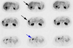
Because of damage to cardiomyocytes and the contractile apparatus, contractile reserve may be observed less frequently in hibernating than in stunned myocardium. The aim of this study was to assess the presence of contractile reserve in response to dobutamine infusion in a large group of patients with stunned and hibernating myocardium. METHODS: A total of 198 consecutive patients with ischemic cardiomyopathy (left ventricular ejection fraction < or = 40%) underwent resting 2-dimensional echocardiography to assess regional contractile dysfunction. On the basis of assessment of perfusion (with (99m)Tc-tetrofosmin SPECT) and glucose use (with (18)F-FDG SPECT), dysfunctional segments were grouped. Dysfunctional segments with normal perfusion were classified as stunned. Dysfunctional segments with a perfusion defect were classified as hibernating when a perfusion-(18)F-FDG mismatch was present. Dysfunctional segments with a perfusion defect were classified as scar tissue when a perfusion-(18)F-FDG match was present; these segments were subdivided into nontransmural and transmural scars. Contractile reserve was evaluated by dobutamine stress echocardiography. RESULTS: Dobutamine-induced contractile reserve was more frequently found in stunned than in hibernating myocardium (61% vs. 51%, respectively; P < 0.01). Only 14% of the scarred segments improved in wall motion during dobutamine infusion, significantly less than stunned or hibernating myocardium (P < 0.001). Nontransmural scars exhibited contractile reserve more frequently than did transmural scars. CONCLUSION: The progressive reduction of contractile reserve in stunned, hibernating, and scarred myocardium supports the hypothesis that stunning, hibernation, and scarring are not circumscript pathophysiologic entities but represent gradual ultrastructural damage on the myocyte level.
