(18)F-FDG accumulation in
atherosclerotic plaques: immunohistochemical and PET imaging study.
Ogawa M, Ishino S, Mukai T, Asano D, Teramoto N, Watabe H, Kudomi N, Shiomi M,
Magata Y, Iida H, Saji H.
The rupture of atherosclerotic plaques and the subsequent formation of thrombi
are the main factors responsible for myocardial and cerebral infarctions. Thus,
the detection of vulnerable plaques in atherosclerotic lesions is a desirable
goal, and attempts to image these plaques with (18)F-FDG have been made. In the
present study, the relationship between the accumulation of (18)F-FDG and the
biologic characteristics of atherosclerotic lesions was investigated.
Furthermore, PET imaging of vulnerable plaques was performed with an animal
model of atherosclerosis, Watanabe heritable hyperlipidemic (WHHL) rabbits.
METHODS: WHHL (n = 11) and control (n = 3) rabbits were injected intravenously
with (18)F-FDG, and the thoracic and abdominal aortas were removed 4 h after
injection. The accumulated radioactivity was measured, and the number of
macrophages and the intimal area were investigated by examination of stained
sections. PET and CT images were also acquired at 210 min after injection of the
radiotracer. RESULTS: (18)F-FDG accumulated to a significantly higher level in
the aortas of the WHHL rabbits (mean +/- SD differential uptake ratio [DUR],
1.47 +/- 0.90) than in those of the control rabbits (DUR, 0.44 +/- 0.15); DUR
was calculated as (tissue activity/tissue weight)/(injected radiotracer
activity/animal body weight), with activities given in becquerels and weights
given in kilograms. (18)F-FDG uptake and the number of macrophages were strongly
correlated in the atherosclerotic lesions of the WHHL rabbits (R = 0.81). In the
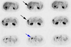
PET analysis, intense (18)F-FDG radioactivity was detected in the aortas of the
WHHL rabbits, whereas little radioactivity was seen in the control rabbits.
CONCLUSION: The results suggest that macrophages are responsible for the
accumulation of (18)F-FDG in atherosclerotic lesions. Because vulnerable plaques
are rich in macrophages, (18)F-FDG imaging should be useful for the selective
detection of such plaques.
PET > PET tumor imaging > General
Latest in PET
PET > PET tumor imaging > Neuroendocrine tumors
April 14, 2021
PET > PET tumor imaging > Head and Neck Tumors
March 12, 2018
PET > General > Images
January 23, 2018
PET > PET tumor imaging > Prostate cancer
January 20, 2016
