Salaun PY, Grewal RK, Dodamane I, Yeung HW, Larson SM, Strauss HW.
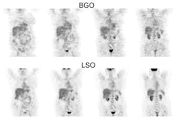
The concentration of (18)F-FDG in the gastroesophageal junction (GEJ) and gastric antrum (GA) varies significantly from patient to patient. To document the reference range of uptake in patients, we reviewed the (18)F-FDG PET scans of patients with no documented gastroesophageal disease. METHODS: The medical records of patients undergoing PET/CT were reviewed. Patients with known gastric, pancreatic, or liver pathology were excluded. The peak standardized uptake value (SUV) for the GEJ and GA were measured in the remaining patients. The clinical record was also reviewed for gastroesophageal reflux disease (GERD) and previous chemotherapy. RESULTS: A total of 763 patients met the inclusion criteria (388 male and 375 female; mean age +/- SD, 57.4 +/- 17 y; range, 15-95 y). Images were recorded 68.2 +/- 11.8 min after injection of 558.7 +/- 35.1 MBq of (18)F-FDG. PET/CT was performed on a Discovery LS scanner for 373 patients and on a Biograph scanner for 390. The maximum SUV was less than 4 in 94.4% of patients. GEJ SUV measurements on the Discovery LS and on the Biograph did not significantly differ. During the 6 mo before the scan, 515 patients received no antineoplastic chemotherapy. Of the remaining 248, 137 received chemotherapy within 1 mo before the scan; 65, between 1 and 3 mo before the scan; and 46, between 3 and 6 mo before the scan. No significant differences were found between groups. GERD was documented in the records of 75 patients. Only 58 of these patients were treated with an antacid regimen. In 552 patients, GERD was not known to be present nor was antacid treatment used. An additional 136 patients had antacid treatment without specified gastric symptomatology. Patients with a history of GERD had a slightly higher but not statistically significant SUV peak in the stomach and particularly in the GEJ, except when compared with the group without associated antacid treatment (P = 0.049). CONCLUSION: In patients without a specific history of esophagogastric disease, a gastroesophageal maximum SUV less than 4 is usually not associated with gastroesophageal neoplasia.


