Truong MT, Erasmus JJ, Munden RF, Marom EM, Sabloff BS, Gladish GW, Podoloff DA, Macapinlac HA.
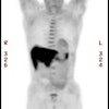
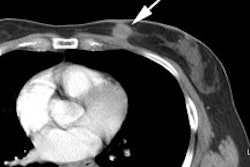
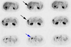
OBJECTIVE: A potential source of false-positive FDG PET interpretations in oncologic imaging is FDG uptake in brown fat. The purpose of this study was to determine the prevalence, location, and appearance of hypermetabolic brown fat in the mediastinum. MATERIALS AND METHODS: All PET/CT scans obtained at our cancer institution from August to October 2003 were retrospectively reviewed for increased FDG uptake in the mediastinum localized to fat on CT. The following features were recorded: location, appearance, maximal standard uptake value (SUV(max)) of hypermetabolic mediastinal brown fat, and presence of extramediastinal brown fat. RESULTS: PET/CT scans were obtained in 845 oncologic patients. Fifteen patients (1.8%) with focal hypermetabolic mediastinal brown fat were identified: nine women and two men (age range, 27-79; mean, 55.1 years) and four children (age range, 5-16 years; mean, 10 years). Hypermetabolic mediastinal brown fat (mean SUV(max), 5.7) was more common in children (4/8) than in adults (11/837) and more common in women (9/372) than in men (2/465). Foci of hypermetabolic brown fat were localized to the paratracheal, paraesophageal, prevascular, and pericardial regions; interatrial septum; and azygoesophageal recess. Five patients had focal hypermetabolic brown fat isolated to the mediastinum. Ten patients also had extramediastinal hypermetabolic brown fat in the neck, thorax, and abdomen. There was no difference in the body weight (p = 0.876) or body mass index (p = 0.538) of patients with hypermetabolic brown fat compared with age- and sex-matched control subjects. CONCLUSION: Hypermetabolic brown fat can be localized to the mediastinum and manifests as focal increased FDG uptake. Knowledge of this potential pitfall and precise localization with fusion PET/CT are important in preventing misinterpretation as malignancy.
