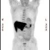
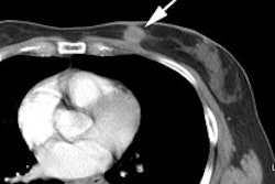
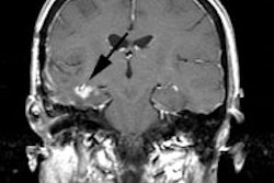
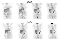
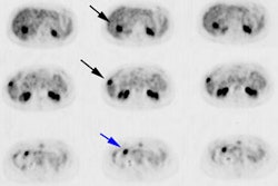
18F-FDG PET of patients with Hurthle cell carcinoma.
Lowe VJ, Mullan BP, Hay ID, McIver B, Kasperbauer JL.
Hurthle cell carcinoma is an uncommon differentiated thyroid cancer characterized by an aggressive clinical course and low avidity for (131)I. Treatment usually involves an aggressive surgical approach often combined with (131)I. (18)F-FDG PET has been helpful in the staging and evaluation of many types of aggressive malignancy. No reports to date have described the utility of PET in a series of patients with Hurthle cell cancer. We reviewed our experience with (18)F-FDG PET in the care of patients with Hurthle cell carcinoma to determine the likelihood of uptake in these cancers and the effect of (18)F-FDG PET on patient care. METHODS: Patients with Hurthle cell cancer who were seen between June 2000 and April 2002 and were imaged with (18)F-FDG PET were included. Imaging and clinical data were reviewed. PET results were compared with the results of anatomic imaging (CT, sonography, or MRI) and (131)I imaging when performed. Patient charts were reviewed to identify any change in management that resulted from the (18)F-FDG PET findings. RESULTS: Fourteen (18)F-FDG PET scans of 12 patients were obtained in the time frame indicated. All patients had documented Hurthle cell carcinoma. PET showed intense (18)F-FDG uptake in all known Hurthle cell cancer lesions but one. PET showed disease not identified by other imaging methods in 7 of the 14 PET scans. PET identified distant metastatic disease (5) or local disease (2) that was more extensive than otherwise demonstrated. In 7 of the 14 scans, the information provided by PET was used to guide or change therapy. CONCLUSION: Hurthle cell carcinoma demonstrates intense uptake on (18)F-FDG PET images. PET improves disease detection and disease management in patients with Hurthle cell carcinoma relative to anatomic or iodine imaging. (18)F-FDG PET should be recommended for the evaluation and clinical management of patients with Hurthle cell carcinoma.
