Introduction
Before the advent of snowboarding, fractures of the lateral process of the talus (LPT) were rare injuries, usually associated with severe trauma or a direct crush injury (Journal of Bone and Joint Surgery, 1965, Vol.47A:6).
Since the early 1980s, however, the incidence of lateral talar fractures has increased with the fast-rising popularity of the sport, and the association of the two has become well recognized (Therapeutische Umschau, December 2000, Vol.57:12, pp. 756-759).
The largest epidemiological study to date reports that LPT fractures account for 15% of all snowboarding injuries (American Journal of Sports Medicine, March-April 1998, Vol.26:2, pp. 271-277).
Clinical history
Twenty-five-year-old man who injured his ankle while doing aerial acrobatics on his snowboard.
Physical exam and radiographic findings
The symptoms are similar to an ankle sprain, so the diagnosis of a fracture is often delayed. Compounding the clinical difficulty of recognizing this injury, routine radiographs frequently do not demonstrate LPT fractures; they are missed 40%-50% of the time on an initial exam (Journal of Orthopedic Trauma, August1994, Vol.8:4, pp.332-337).
Plain-film examination also tends to underestimate the extent of injury and the degree of displacement of the fracture fragments.
 |
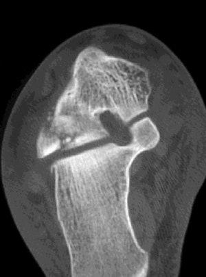
| Axial CT image demonstrates comminuted fracture of the lateral process of the thalus that extends into the posterior talocalcaneal joint. |
 |
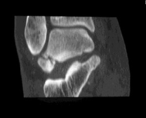
| Coronal reconstruction CT image demonstrates extension of the fracture of the lateral process of the talus into the inferior talofibular joint. |
 |
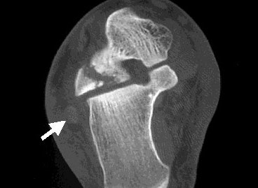
| Axial CT scan shows that the largest fracture fragment is displaced 3-4 mm posterolaterally. The peroneal tendons (white arrow) do not appear to be affected by the fracture fragments. Images courtesy of Dr. Douglas Beall. |
Discussion
Processus lateralis tali fractures are thought to result from inversion in dorsiflexion (causing a compression force on the lateral talus) and have been classified into three types: Type 1 is a nonarticular chip fracture that may be treated conservatively. Type 2 fracture has a single large fragment and involves the talofibular or subtalar joint. Type 3 fracture is comminuted and involves both joints (Journal of Bone and Joint Surgery, 1965, Vol.47A:6).
Prompt recognition of an LPT fracture is important, given that approximately 25% of these fractures will develop osteonecrosis. Malunion and post-traumatic osteoarthritis are also frequent sequelae that may result from a delayed diagnosis.
These complications are not trivial. The talus is one of the primary weight-bearing bones in the hindfoot, and the symptoms resulting from these complications can be severe and debilitating. Any snowboarder with persistent and/or severe acute ankle pain, limitation of ankle movement, or who fails to respond to conservative therapy should be evaluated for an LPT fracture.
Due to the limitations of routine radiographs in LPT fractures, CT scanning should be used in all patients suspected of having this type of fracture. CT can be used to determine the size or degree of comminution of the fractured process, as well as to demonstrate the extent of subtalar involvement, any detectable tendon pathology, and other associated fractures (Therapeutische Umschau, December 2000, Vol.57:12, pp. 756-759).
Many other associated fractures of the hindfoot and midfoot can also be subtle, and difficult to evaluate by plain-film radiography. However, they can also be detected by thin-section spiral CT of the foot and ankle.
Evaluation of the anatomy of the fracture is very important for determining the appropriate treatment and assessing the need for surgery. Comminuted fractures, fractures that extend into joints, and fracture fragments with displacements of 2 mm or more are all associated with more complications and a higher rate of nonunion.
Most physicians agree that nondisplaced fractures are most appropriately treated conservatively by applying a cast and immobilizing the foot. Displaced fractures require surgical treatment, and single large fragments are reduced and internally fixed, while smaller comminuted fragments may need excision.
Generally, after 48-72 hours of bed rest, the patient is allowed to ambulate with only limited partial weight bearing on the affected extremity. The time allowed for this fracture to heal is similar to other types of fractures, and most young healthy individuals have adequate healing after approximately 6 weeks.
Conclusion
The increased popularity of snowboarding has brought about an increase in the prevalence of fractures of the lateral process of the talus. This type of fracture now has an established association with snowboarding, and accounts for a significant portion of ankle injuries seen in individuals participating in this sport.
This fracture may mimic a lateral ankle sprain and is easily missed on plain-film radiographs of the ankle. Due to the relatively high complication rates and to the significant disability of an unrecognized LPT fracture, CT should be used to evaluate all snowboarders suspected of having this type of injury.
Given the difficulty of making this diagnosis, both primary care physicians and specialists should be aware of the association of this fracture with snowboarding; they should maintain a low threshold for ordering cross-sectional imaging for these patients.
By Dr. Douglas P. BeallAuntMinnie.com contributing writer
February 24, 2002
Dr. Beall is a staff radiologist in the musculoskeletal division, department of radiology at Wilford Hall Medical Center, Lackland Air Force Base, TX. He also is an assistant professor of radiology, department of radiology and nuclear medicine, Uniformed Services Health University in Bethesda, MD.
The opinions and assertions herein are the private views of the author and are not to be construed as official or as reflecting the view of the Department of the U.S. Air Force, U.S. Department of Defense, or the U.S. government.
Copyright © 2002 AuntMinnie.com


