Panoramic radiography's potential role in assessing atherosclerotic risk is highlighted by a new study that shows an association between findings of periodontal bone loss and carotid plaque thickness.
Using x-rays to find bone loss is quicker and less invasive than clinical methods for gauging chronic periodontitis, an infection that has been associated with stroke and subclinical atherosclerosis in a series of studies.
The report from the Oral Infections and Vascular Disease Epidemiology Study (INVEST) is the latest delineating the increased risk of stroke faced by patients with chronic infections (Stroke, March 2005, Vol. 36:3, pp. 561-566).
Some researchers have theorized that chronic infections may induce systemic inflammation or autoimmunity that leads to atherosclerosis.
Investigators on the latest report include lead author Steven P. Engebretson, DMD, and senior author Dr. Moise Desvarieux, from the periodontics and epidemiology divisions, respectively, at New York City's Columbia University.
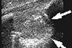
The INVEST researchers used x-rays to measure periodontal bone loss, then compared those findings to duplex Doppler ultrasound of the carotid arteries in 203 study participants.
Panoramic radiographs were taken by dental school personnel, then read and scored by a trained examiner, the authors wrote. The severity of bone loss was gauged by the percentage missing at the mesial and distal surfaces of each tooth.
Ultrasonography was performed by certified sonographers using a Logiq 700 system (GE Healthcare, Chalfont St. Giles, U.K.) equipped with a multifrequency 9/13-MHz linear-array transducer. The presence of atherosclerotic plaque was defined as an area of focal wall thickening.
The prevalence of carotid artery plaque in the study group was 57%, the authors reported
After adjusting for age, hypertension, history of cardiovascular disease, diabetes, smoking, high-density lipoprotein (HDL), and low-density lipoprotein (LDL), a finding of severe periodontal bone loss was significantly associated with carotid artery plaque, with an odds ratio of 3.64.
Additional analyses that looked at the impact of education and race or ethnicity did not change the results, the researchers reported.
Their report reflects the first cross-sectional study to investigate the association between carotid artery plaque and periodontal bone loss, and the first to use radiographs to assess chronic periodontitis in a research study of subclinical atherosclerosis.
The authors suggested that panoramic x-rays might be the diagnostic tool of choice for their future investigations.
"Radiographic measurement of alveolar bone is an accurate method for assessment of periodontitis and compares favorably with clinical measures of periodontitis," they wrote. "The panoramic radiograph can be performed in a few minutes in relative patient comfort compared with 20 to 30 minutes for a complete clinical oral evaluation."
Oral radiographs might even be useful in screening for carotid artery calcifications, the authors wrote, noting an earlier study of 83 patients that found a high degree of correlation between the periodontitis and calcifications visualized just on panoramic x-rays.
"In summary, this study adds to the growing body of evidence that chronic periodontitis (CP) is an important and independent risk factor for atherosclerosis," the authors concluded. "Panoramic oral radiographs provide an efficient means to assess CP in studies of atherosclerosis risk."
By Tracie L. Thompson
AuntMinnie.com staff writer
April 1, 2005
Related Reading
Skin cholesterol levels mirror levels in carotid arteries, March 8, 2005
Periodontal pathogens linked to atherosclerosis, February 10, 2005
MRI for carotid artery wall thickness measures up to US gold standard, February 3, 2005
Multidetector CT angiography identifies stable carotid plaques, January 12, 2005
MDCT advances oral and maxillofacial surgery practice, November 22, 2004
Copyright © 2005 AuntMinnie.com


