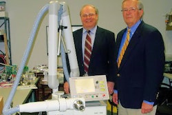
Electromagnetic (EM) imaging can boost the contrast between normal breast tissue and abnormal findings by as much as 200%, according to the results of a study by Dartmouth University-based researchers. But their prototype scanners still require a fair amount of fine-tuning before they'll be useful in a high-throughput clinical setting.
The electromagnetic properties of breast tissue offer high contrast for depicting malignancies when electrical impedance spectroscopy (EIS) is used, wrote Dr. Steven Poplack and colleagues. Additionally, microwave impedance spectroscopy (MIS) and near-infrared spectral tomography (NIR) have the potential to discriminate between breast cancer and benign conditions following an abnormal standard mammogram.
The study was "our initial attempt to exploit a multimodality framework for enhancing breast cancer diagnosis," said Poplack, who is from the department of radiology at Dartmouth-Hitchcock Medical Center in Lebanon, NH. His co-authors are from the Lebanon-based medical school and the Norris Cotton Cancer Center, as well as the Thayer School of Engineering in Hanover, NH (Radiology, March 30, 2007).
Ninety-seven women, either with suspicious findings (BI-RADS 4-5) or negative findings (BI-RADS 1) on conventional breast imaging (mammography and/or sonography), made up the abnormal group and the control group, respectively.
The subjects in the abnormal group underwent EM imaging a mean of 2.5 days before percutaneous breast biopsy. Normal subjects had EM exams a mean of 78 days after screening. Thirty-one participants with malignant biopsy results and two with atypical results had surgical resection.
EM imaging was done using previously described techniques. The patient was in a prone position and the imaging array circumscribed the pendant breast (IEEE Transactions on Biomedical Engineering, January 2000, Vol. 47:1, pp. 49-58; Review of Scientific Instruments, July 2004, Vol. 75:7, pp. 2305-2313, and March 2001, Vol. 73:2, pp. 1817-1824).
A region of interest (ROI) analysis was done to compare EM image properties to conventional imaging findings and pathologic parameters. Receiver operating characteristic (ROC) curves were compared for abnormal and normal subjects.
Of the 31 women in the abnormal group, 32% had breast cancer, with nine cases of ductal carcinoma in situ (DCIS). Forty-nine women had fibrocystic disease, according to the results.
The EM imaging results were as follows:
Among 62 women in the abnormal group who underwent EIS, the findings were classified as fibrocystic disease in 32 cases and malignancies in 16. The area under the curve (AUC) was estimated at 0.78 in this group. The AUC for cancer-noncancer grouping was 0.66 and 0.59 in the cancer-benign lesion grouping.
Among 80 women in the abnormal group who had MIS, there were 26 malignancies and 41 sites of fibrocystic disease. The AUC was estimated at 0.80. The AUC for the cancer-noncancer grouping was 0.83 and cancer-benign lesion was 0.89.
Among the 58 women in the abnormal group who underwent NIR, there were 18 malignancies and 31 fibrocystic disease sites. The AUC for the cancer-noncancer group grouping was 0.82 and 0.76 for the cancer-benign lesion grouping.
"EM property indicators with the highest contrast ratios for cancer appear to be electrical permittivity at EIS (mean contrast ratio, 1.6), electrical conductivity at MIS (mean contrast ratio for lesions > 1 cm in size, 2.0), and total hemoglobin concentration at NIR (mean contrast ration for lesions > 6 mm in size, 1.5)," the authors explained. "EIS … appears to help in the discrimination between normal and abnormal breast tissue."
However, they cautioned that EIS did not seem to aid in differentiating cancer from other abnormal pathology.
MIS results provided better conductivity for great contrast and discrimination, and their NIR results approached those reported in a breast MR study, they stated (Journal of the American Medical Association, December 8, 2004, Vol. 292:22, pp. 2735-2742).
Ultimately, the group reported a mean increase in image contrast of 150% to 200% between abnormal and normal breast tissue. Combining EIS, MIS, and NIR did incrementally increase the diagnostic value of EM, they concluded, although the exam time of multimodality EM is currently too long for use in practice. This, and other technical limitations, still need to be overcome, they added.
By Shalmali Pal
AuntMinnie.com staff writer
April 10, 2007
Related Reading
Electromagnetic tracking system can guide flexible bronchoscopy, July 20, 2005
Breast biopsy costs big bucks, but so does cancer screening, January 19, 2005
Copyright © 2007 AuntMinnie.com


