Dear AuntMinnie Member,
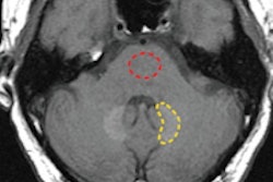
A disturbing new study published this month in Radiology indicates that traces of gadolinium from MRI contrast agents may persist for years in the brains of patients who have received scans.
Researchers from the Mayo Clinic in Rochester, MN, found traces of gadolinium in the brain tissue of 13 patients who received MRI scans over a 14-year period. The traces were found in all patients who had received gadolinium contrast -- and even in individuals who got as few as four doses of contrast.
In 2006, gadolinium was implicated in the development of nephrogenic systemic fibrosis (NSF), a severely debilitating and sometimes fatal disease, but NSF incidence dropped dramatically after imaging facilities adopted protocols restricting its use in patients with compromised renal function.
What's the clinical significance of the new findings? The Mayo researchers say they aren't sure, although they noted that the patients with gadolinium traces had normal renal function. Learn more by clicking here.
While you're in our MRI Community, check out this story on a new technique that uses MRI to detect sugar shed by malignant lesions, offering a potentially new way to detect cancer.
Risks of skipping mammo screening
Meanwhile, in our Women's Imaging Community, we're covering a new study published in the American Journal of Roentgenology that highlights the risks of skipping annual mammography exams.
Researchers from Wisconsin reviewed the mammography screening records over five years for 1,400 women who had received a diagnosis of breast cancer. They found that women who had missed two annual mammography screenings had nearly double the all-cause mortality as women who didn't miss any screenings -- and mortality rose with the number of missed exams.
The findings are sure to fuel the debate over mammography screening, especially given the ongoing fight over whether screening should be performed annually or every two years. Learn more by clicking here, or go to women.auntminnie.com.
US first for female pelvis
Finally, a new study is recommending that ultrasound be used first for female pelvic imaging instead of MRI or CT. Researchers from Harvard Medical School believe that recent developments in ultrasound technology make it well-suited for the application, and it can avoid more expensive tests, some of which involve radiation. Read more by clicking here for an article in our Ultrasound Community.


