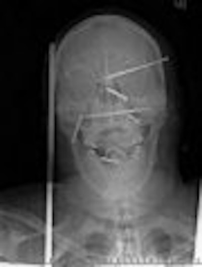
When the patient arrived at Providence Holy Cross Medical Center in Mission Hills, CA, it was clear he'd need more than an x-ray. But plain films were able to answer an immediate question: How many nails had been driven into his head?
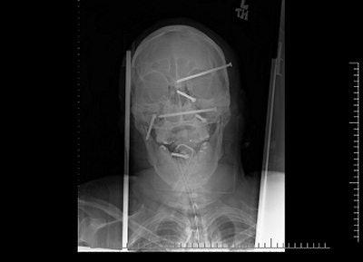
Six nails, as it turned out, could be seen by anyone who saw the x-ray images released to the media earlier this year, after construction worker Isidro Mejia's impressive recovery from a horrific nail-gun accident.
Now, right here on AuntMinnie.com, you can also see the 3D CT images that proved most useful for Mejia's treatment. Not that the radiographs (below) weren't revealing.
 |
| Image courtesy of Providence Holy Cross Medical Center. |
"Nobody knew the extent of the injury until the first x-ray," said Dr. Rafael Quinonez, the neurosurgeon who operated on Mejia. "Nothing was sticking out."
"On physical exam you didn't see most of these," agreed staff radiologist Dr. Stephen Greenberg. "You'd see puncture wounds, you'd see blood, but you wouldn't really know exactly where the nails were."
Mejia was working on the roof of an unfinished home on April 19 when he fell down onto a co-worker who was using a high-powered nail driver. The men grabbed at each other to keep from falling further; meanwhile the loose nail gun kept discharging.
Initial x-rays showed there were six 3.5-inch nails in Mejia's head, and that his jaw bones remained intact. He immediately underwent a CT scan, with initial images showing significant metal artifact, as expected (images A-B below).
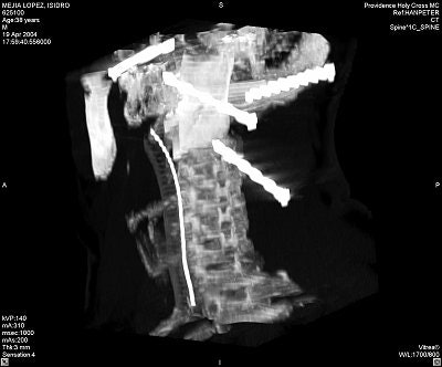 |
Image A.
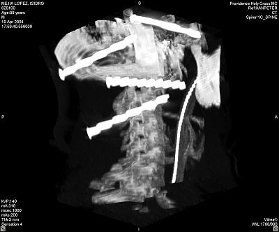 |
"Basically you could only visualize on hard-bone algorithm because of the nails," noted Greenberg. "You had very little ability to tell how much bleeding the nails had caused, and certainly you had no way to tell whether there had been injury to a specific arterial structure because there was no way to get any soft-tissue detail."
But the CT provided enough information for Quinonez to allow the patient to stabilize for a few days, when a conventional angiogram could be performed.
"The angiogram basically just defined how lucky (Mejia) was, and that all the vessels were basically where they were supposed to be," Greenberg said. "When you had several views you could see the vessels were taking a normal course and the nails had managed to miss them all."
Quinonez was able to remove the nails based on the angiogram and 3D CT reconstructions that showed the location of the nail heads -- most critically, the head of a nail that had entered anteriorly through the base of the skull into the face and that now rested medial to the mandibular condyle (images C-E below).
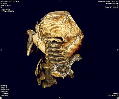 |
Image C.
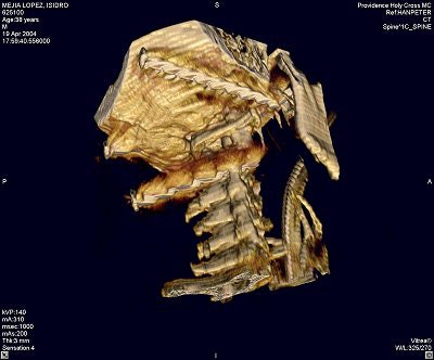 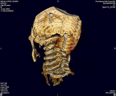 |
"There was concern as to (a nail's) relationship to several branches of the carotid artery," noted Greenberg. "The nail was going to be taken out from the left side of his neck, but the sharp end was on the right side, where the surgeon was not going to have access if something was going to start bleeding."
Needless to say, MR imaging wasn't an option with this patient. Still, Greenberg was able to employ the modality for some twisted hospital humor.
"I made a joke, at the time that the patient was first admitted, that I offered to put the patient into the magnet in an attempt to remove the nails and save him the surgery," said Greenberg, "but that I didn't have a CPT billing code for nail removal."
By Tracie L. ThompsonAuntMinnie.com staff writer
July 13, 2004
Related Reading
CT can reduce unneeded laparotomy for patients with gunshot wounds, May 21, 2004
Use of pediatric head CT growing steadily in emergency departments, May 4, 2004
Imaging in Trauma and Critical Care, January 20, 2004
3-D and the future of imaging, November 3, 2003
Copyright © 2004 AuntMinnie.com


