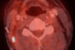
Hot on the tail of the first mouse MR colonography study, investigators in Wisconsin have established a technique to perform microCT virtual colonoscopy in 20 murine subjects from Madison.
"The study was initiated to evaluate the efficacy of negative contrast-enhanced microcomputed tomography (microCT) for the noninvasive detection of tumors in living mice," wrote Dr. Perry Pickhardt; Richard Halberg, Ph.D.; Dr. Andrew Taylor; and colleagues from the University of Wisconsin Medical School and the McArdle Laboratory for Cancer Research in Madison, WI.
Mouse colonography is harder than it might seem. Imaging systems used for human studies generally lack the high spatial resolution needed to image small animals, necessitating the development of dedicated high-resolution systems. The x-ray microCT scanner (Micro-CAT I, ImTek, Knoxville, TN) used in the study can achieve spatial resolution on the order of 20 µm, the researchers wrote in the Proceedings of the National Academy of Sciences (March 1, 2005, Vol. 102:9, pp. 3419-3422).
Still, fewer studies have been conducted with microCT than with other micromodalities, and even fewer microCT studies have been done in soft-tissue applications such as the colon, the authors wrote, due in part to the rapid clearance of contrast materials compared to the long imaging times, 20 minutes in the present study, that are required.
Colonic polyps and tumors were created in the mice by introducing a congenic strain based on the Min allele of the Apc gene modified from the C57BL/6J background.
Before imaging, the 20 congenic mice (17-22 g body weight) were fed a diet of fresh vegetables, sunflower nuts, and water for two days, followed by cherry-flavored NuLytely (Braintree Scientific, Braintree, MA) for 16 hours before scanning. Following anesthetization (pentobarbital, 0.006 mg/g body weight) and a corn-oil enema (1-1.5 mL), the animals were imaged using microCT. No gating or contrast administration was used, the authors noted.
Images were acquired at 43 kVp, 410 µA, and 390 steps, with a 20-minute scan duration. Image data were reconstructed at 200-µm spatial resolution (256 x 256 x 256 voxels). Reconstruction of the image data was performed using a Shepp-Logan filter with back projection and no beam-hardening correction, the group wrote.
Amira visualization software (Version 3.1, TGS, San Diego) was used to display the data as axial, sagittal, and coronal cross-sectional images in 2D, and the data were reviewed by two experienced gastrointestinal radiologists who were unaware of the pathological results.
Polyps were distinguished from fecal pellets on a five-point scale, with 1 representing "definitely not a tumor." The animals were killed immediately after imaging, and gross pathological inspection of the excised mouse colon served as the gold standard.
In all, 41 tumors were seen at pathology, including two lesions measuring 5 mm, seven at 4 mm, 11 at 3 mm, 10 at 2 mm, and 11 smaller than 2 mm (mean 3.0 mm). Seventeen mice had a polyp measuring 2-5 mm.
"The ability to detect endoluminal lesions was significantly enhanced in low-density bowel-prepped mice relative to normal chow-fed mice," the authors wrote. (Chow-fed rodents in previous attempts to develop the protocol had been unsuccessful.)
"Although fecal pellets are visible ... by virtue of their increased density in unprepped mice, it remains virtually impossible to detect intestinal lesions in these animals. Negative contrast enhancement of the bowel, however, affords a much higher contrast differential in the lumen, thus enhancing the conspicuity of lesions within the lumen."
For lesions larger than 2 mm, the pooled per-polyp sensitivity was 93.3% (56/60); pooled per-mouse sensitivity was 97.1%. The pooled sensitivity for distinguishing mouse pellets from tumors was 98.5% (65/66), the authors wrote. And at the 2-mm threshold, per-mouse sensitivity for excluding polyps was 100% (3/3). The combined area under the curve from ROC curve analysis was 0.810 ± 0.038 (95% confidence interval, 0.730-0.890).
"Although soft-tissue tumor detection can benefit greatly by using IV contrast agents in human CT, evaluation of soft-tissue tumor models by microCT is severely hampered by the rapid clearance of conventional contrast agents relative to the long imaging times that are required in microCT," the group wrote. "The use of targeted contrast agents with a much longer organ or blood pool half-life represents one solution. For the case of colorectal tumors, however, protrusion into the bowel lumen provides an intrinsic contrast gradient that makes IV contrast unnecessary for detection."
Although the sample size was small, the authors said they believed proof of concept had been established.
"Accurate scanning of live mice with microCT indicates that longitudinal monitoring of tumor growth and response to therapy should now be feasible," they wrote. "The result of future studies with microCT colonography could serve as a direct preclinical bridge to studies involving human studies."
By Eric Barnes
AuntMinnie.com staff writer
March 16, 2005
Related Reading
Mutant survivin gene delivery inhibits colon cancer in mice, February 23, 2005
TGF-beta suppresses colon cancer progression, November 22, 2004
MYH gene mutation tied to colorectal cancer, November 9, 2004
MR colonography finds most tiny rat lesions, October 8, 2004
Copyright © 2005 AuntMinnie.com


