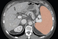
(Radiology Review) Among the splenic indexes used for evaluating splenomegaly, a simple CT measurement of the splenic length and the relationship between the inferior spleen and the inferior third of the left kidney are strong predictors of splenomegaly, according to researchers from the Catholic University Hospital in Brazil.
Splenic length (sensitivity 85%, specificity 89%) and width (sensitivity 82%, specificity 81%) demonstrated the best correlation to splenic volume, the researchers reported in the American Journal of Roentgenology (May 2005, Vol. 184:5, pp. 1510-1513).
The study included 249 adult patients whose splenic length, width, and thickness were measured on CT. From these measurements, splenic volume and the relationship between the inferior spleen and surrounding organs, such as left lobe of liver and left kidney, were evaluated. Sixty-two patients (24.8%) patients had splenomegaly.
CT was performed using a Tomoscan AV single-detector helical scanner (Philips Medical Systems, Andover, MA). During a single breath-hold, scans were obtained using a 7-mm section thickness, 7-mm reconstructed section thickness, and 7-mm reconstruction interval.
A summation-of-volumes technique using an EasyVision workstation (Philips Medical Systems) was used to calculate splenic volume. Each splenic outline was measured to calculate the enclosed area, then the area was multiplied by the section thickness to derive the volume of that axial section. Summation of each section volume gave a total splenic volume.
Dr. Alexandre S. Bezerra and colleagues used an established upper limit of normal for splenic volume (314.5 cm3), and derived a maximum spleen length of 9.76 cm and a maximum spleen width of 11 cm from the linear regression equation. Also, they determined that when the spleen reached or extended below the lower third of the left kidney, this was highly specific for splenomegaly despite a low sensitivity (sensitivity 20%, specificity 93%).
The study validated the use of a simple splenic length measurement for splenic CT volume. When splenic length exceeds 10 cm, a diagnosis of splenomegaly should be made. This clinically practical, accurate, simple measurement replaces multiple-measurement, time-consuming methods, and allows daily routine follow-up, the authors reported.
"Determination of Splenomegaly by CT: Is There a Place for a Single Measurement?"
Alexandre S. Bezerra, et al
Universidade Católica de Brasília-Campus I
Curso de Medicina
EPCT QS 7 Lote 1
Taguatinga, Distrito Federal, Brazil, 71966-700
AJR 2005 (May); 184:1510-1513
By Radiology Review
May 11, 2005
Copyright © 2005 AuntMinnie.com


