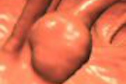
Ask any gastroenterologist who performs colonoscopy -- probably the easiest way to miss a polyp is to fly right past it. The problem may occur even more frequently in 3D virtual colonoscopy, where polyps are missed when the radiologist doesn't steer his virtual colonoscope to the lesion's hiding spot.
But a new software application aims to eliminate the problem by highlighting areas that were missed during the first two endoluminal fly-throughs, so they can be examined in a third look.
Primary 3D endoluminal viewing differs from primary 2D reading of virtual colonoscopy data, said Dr. Anno Graser from the University of Munich-Grosshadern in Germany at the 2006 European Congress of Radiology (ECR) in Vienna.
While primary 3D offers an anatomical view with "long contact time eye to lesion," it requires two fly-throughs from the rectum to the cecum and back again, Graser said. Primary 2D viewing reduces the time that a polyp is visible, but requires only one scroll-through.
In 3D reading there is a "key problem with radiologists not evaluating the entire colonic mucosa," he said. As a result, areas behind haustral folds often remain unseen.
In fact, a study by Yasumoto et al found that unidirectional fly-through detects fewer lesions than bidirectional fly-though: 78.1 versus 84.4%, respectively (American Journal of Roentgenology, January 2006, Vol. 186:1, pp. 85-90).
Graser and colleagues sought to assess the percentage of colonic surface not seen during fly-throughs using a prototype workstation (Siemens Leonardo Colon, Siemens Medical Solutions, Erlangen, Germany) equipped with an unseen areas visualization tool. The software has since received 510(k) approval as a component of syngo Colonography in the syngo MultiModality Workplace, according to Siemens.
The study included 40 datasets with different patient positions (20 prone, 20 supine), randomly selected from a database and examined on the workstation. The CT data had been acquired on a 64-slice scanner (Somatom Sensation 64, Siemens Medical Solutions) using a full bowel prep but no fecal tagging.
"We performed a unidirectional fly-through from the rectum to the cecum, and then measured the percentage of the colonic surface that had been visualized. In a second step we went from cecum to rectum and again measured the area that had been visualized," Graser said.
After a single fly-through from rectum to cecum, only 71.7% to 88.2% (mean, 81.3%) of the colonic surface had been visualized. A second fly-through increased these values to 91.4% to 98.8% (mean, 96.2%). A significantly larger percentage of the colonic surface was visualized after bidirectional fly-through (p < 0.001, t test), Graser reported. Still there were areas that were unseen.
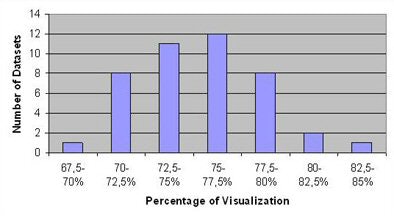 |
| Above, following unidirectional fly-through: 79.4 ± 4.0% of colonic mucosa is visualized (69.5% to 84.5%). Bottom, after bidirectional fly-through: 96.2 ± 2.3% of colonic mucosa visualized (91.4% to 98.8%). The percentage increased to more than 99% after unseen areas are marked for examination. |
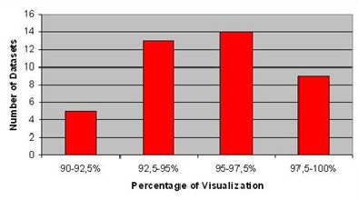 |
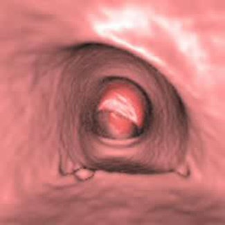
"After the second fly-through, you can use this tool to display unseen areas ... outlined in dark red," he said. "And the 15 largest unseen areas led to visualization of over 99% of the colonic surface in all 40 datasets.
 |
 |
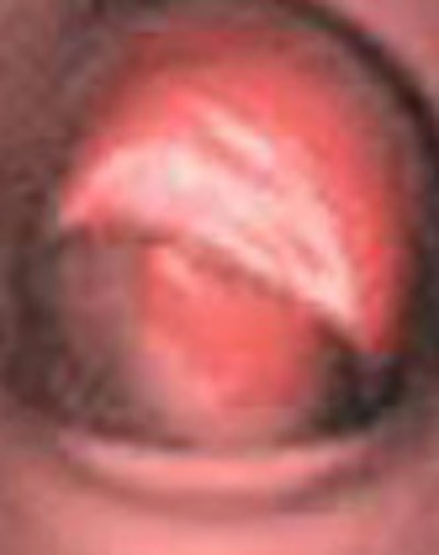
| Video shows a fly-through from the cecum to the rectum with the visualization tool turned on. The radiologist's eye is drawn to the areas in red indicating portions of the colonic surface that have not been visualized. Images, video, and charts courtesy of Dr. Anno Graser. |
The software tool sorts unseen areas according to their size and displayed them to the reader. If the top 15 areas are examined in a third flight, the remaining unseen patches were smaller than 0.25 cm2 in all cases, Graser reported.
"3D software-based display of unseen areas enables visualization of over 99% of the colonic mucosa," Graser concluded. "Bidirectional fly-through visualizes a significantly larger area of the colonic mucosa, and the color coding of unseen areas on flight from cecum to rectum facilitates evaluation of these areas."
In response to a question from the audience, switching the direction of the original fly-through didn't affect the percentage of coverage, Graser said.
"This is a very important paper because one of the major advantages of virtual colonoscopy over colonoscopy is the ability to look back on yourself," commented session moderator Dr. Alan Freeman from the radiology department at the University of Cambridge in the U.K.
By Eric Barnes
AuntMinnie.com staff writer
June 16, 2006
Related Reading
CAD aids polyp detection in 64-slice study, May 12, 2006
'Filet view' VC software slices reading time, October 6, 2005
VC CAD improves results for readers at all levels, April 7, 2006
New VC reading schemes could solve old problems, October 13, 2005
Cubism shapes the art of virtual colonoscopy, October 6, 2003
Copyright © 2006 AuntMinnie.com


