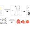
A new study shows that coronary CT angiography (CTA) can be useful for predicting the eventual extent of myocardial dysfunction in patients who have had acute myocardial infarction (AMI), and in those who have undergone reperfusion of an infarct-related artery.
While several previous CTA studies have demonstrated abnormal myocardial enhancement patterns in the presence of AMI, the focus of CTA is lumen visualization for detecting coronary artery stenosis. As for CTA's ability to predict functional recovery at follow-up, a task routinely requested of MRI, the authors wrote in Radiology that they were unaware of any previous reports.
To be sure, previous studies have shown that CTA can demonstrate myocardial abnormalities. "Three types of abnormal myocardial enhancement patterns have been described: (a) hypoenhanced regions on the initial scan and (b) hyperenhanced and (c) hypoenhanced regions on late scans obtained 5-15 minutes after contrast material injection," wrote Dr. Jonathan Lessick and his colleagues from the departments of cardiology and medical imaging at Rambam Health Care Campus and Technion-Israel Institute of Technology, both in Haifa, Israel. "These patterns have been described by using gadolinium-enhanced magnetic resonance (MR) imaging, and the absence of late enhancement (LE) at MR has been verified as strongly predictive of myocardial viability" (Radiology, September 2007, Vol. 244:3, pp. 736-744).
"Thus, the purpose of our study was to prospectively evaluate the sensitivity of myocardial early-perfusion defects (EDs) and LE at multidetector CT following AMI to predict segment myocardial dysfunction and MFR (myocardial functional recovery), by using echocardiography as the reference standard," they wrote.
The study examined 26 patients (25 men, one woman, mean age ± standard deviation, 53 years ± 9) with first AMI. All underwent multidetector CTA on a 16-detector-row scanner (Brilliance, Philips Medical Systems, Andover, MA) between 2002 and 2004. Physicians had established a clinical diagnosis of AMI in all of the patients based on typical clinical symptoms, echocardiographic changes, and elevated creatine phosphokinase or troponin levels.
The mean heart rate was 71 beats per minute and beta-blockers were not used before the scan, the researchers noted. Following power injection of 370 mg/mL of iodinated contrast material (Ultravist, Bayer Schering Pharma, Berlin) at 4 mL/sec, gated CTA images were acquired using 16 x 0.75-mm collimation, 120-140 kVp, 400-600 mAs, with reconstruction intervals of 1 mm. The typical effective dose was 10.5 mSv.
Conebeam corrected reconstructions were performed at 0%, 40% to 50%, and 70% to 80% of the RR interval, and a second scan without additional contrast was performed six minutes after the first at 16 x 1.5-mm collimation, 120 kVp, and 250-250 mAs, with dose modulation used at 70% of the RR interval. For each case the authors examined 16 myocardial segments, paralleling the American Heart Association's 17-segment model without a separate apical segment.
Early echocardiography (within two days of CT) and follow-up at three months were performed and recorded on videotape in all patients using either Sequoia (Siemens Medical Solutions, Erlangen, Germany) or Vivid-3 (GE Healthcare, Chalfont St. Giles, U.K.) equipment.
Invasive coronary angiography was also performed using standard techniques in 23 of the 26 patients, and recorded on a Coroskop Top system (Siemens Medical Solutions). All stenoses were quantitated using electronic calipers, and flow grade was reported.
According to the results, 77% (20/26) patients demonstrated early-perfusion defects and 77% (20/26) had late enhancement. Two patients had neither, demonstrating full left ventricular function at baseline. All early-perfusion defects and late enhancement corresponded with the AMI location determined at conventional angiography and echocardiography.
"Both the presence and the size of a defect were related to the probability of having segment dysfunction," Lessick and colleagues wrote. "The probability of follow-up regional dysfunction for ED increased from 50% for segments in the smallest tertile (ED area, 0.1-0.4 cm²) to 91% for segments in the largest tertile (ED area, 1.0-2.9 cm²), while the likelihood for combined LE increased from 41% for segments in the smallest tertile (LE area, 0.2-0.6 cm²) to 88% for segments in the largest tertile (LE area, 1.5-4.1 cm²)."
In the case of the five occluded arteries, no relationship was found between the presence of ED or LE and myocardial functional recovery. For patent arteries (n = 21), however, the presence of LE had a sensitivity of 73% and specificity of 85% for predicting follow-up segment dysfunction, compared with 57% sensitivity and 90% specificity for ED.
And when baseline segments were abnormal, nonrecovery "was clearly related to the presence and size of segment defect area for both ED (odds ratio: 1.95 [95% confidence interval: 0.9, 4.1] per square centimeter) and LE (odds ratio: 1.85 [95% confidence interval: 1.2, 2.9] per square centimeter)," the group reported. "Segments that recovered had significantly lower prevalence of ED and LE, and if present, were significantly smaller than in segments remaining abnormal (p < 0.05)."
The results show a strong association between the presence and extent of ED and LE and follow-up segmental dysfunction, the team concluded. For patent coronaries, there was also a relationship between the presence and size of ED and LE and the absence of functional recovery.
"The absence of either ED or LE in a particular segment predicts normal function at follow-up, but even segments without any defect had only a 55% chance of recovering, which is much less than that predicted with MR," the authors wrote, adding that more sensitive equipment may eventually identify subtler forms of functional recovery than were found in the patient group.
CTA's ability to predict myocardial functional recovery could be especially important as an ancillary finding in patients who undergo thrombolytic therapy after AMI and CT for coronary artery evaluation, or in patients with acute ischemia with suspected myocardial stunning, they added.
The authors cited as the principal limitations of the study a small study population with potential selection bias, as well as a mean three to four days between AMI and CTA and baseline ultrasound -- because a certain amount of functional improvement may have already occurred during this delay. Misregistration between the modalities would also be expected to add a degree of error to the results.
"Further studies are required to evaluate the clinical value of these preliminary findings in larger patient populations," they wrote.
By Eric Barnes
AuntMinnie.com staff writer
September 7, 2007
Related Reading
High radiation dose with CT angiography warrants caution in children, August 27, 2007
JAMA study: Coronary CTA poses substantial cancer risk in select patients, July 17, 2007
Coronary CTA aids some asymptomatic patients, August 27, 2007
Simple scoring system predicts cardiovascular complications after PCI, June 22, 2007
Copyright © 2007 AuntMinnie.com


