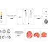
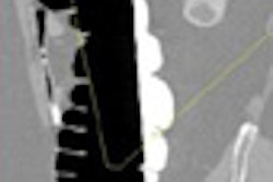
CHICAGO - The use of FDG-PET/CT with patients with inflammatory breast cancer can accurately detect and help treat the deadly disease, according to researchers at the University of Texas M. D. Anderson Cancer Center in Houston.
Inflammatory breast cancer is the most aggressive and deadly form of the disease, and generally affects younger women. About 1% to 5% of all breast cancers in the U.S. are the inflammatory type, and 20% of those cases already involve metastases at presentation. The five-year survival ranges from 20% to 40% of patients, according to Dr. Selin Carkaci, an assistant professor of radiology at M. D. Anderson.
Carkaci demonstrated how the fusion of PET and CT enhances areas where cancer may have spread in women with inflammatory breast cancer in a presentation Tuesday at the RSNA 2007 meeting.
"PET/CT is useful in staging inflammatory breast cancer because it provides information on both the primary disease site, as well as disease involvement throughout the rest of the body," she said. "In addition, PET/CT is also a practical tool for therapeutic planning."
The study at M.D. Anderson's Inflammatory Breast Cancer Clinic and Research Program included 41 patients (mean age 50 years) with newly diagnosed with inflammatory breast cancer. All the women underwent a whole-body FDG-PET/CT exam.
About 20 of the women (49%) had disease that could be easily seen with PET/CT that could not be visualized with CT scanning alone, according to Carkaci. About half the women did not have distant metastases, she said.
The findings were confirmed with through biopsy and supplemental imaging, and there were two false positives and one false negative, for a 95% accuracy rate.
Although the fusion imaging technique had been employed in other cancer disease states, Carkaci said that because of the rarity of the cancer, there have been few studies of PET/CT in inflammatory breast cancer.
"The use of PET/CT will help in treating patients with metastases," said Dr. Joseph Tashjian, president of St. Paul Radiology in Minnesota. He could envision the imaging technique locating a metastasis in the leg of a woman with inflammatory breast cancer. "While we would be giving courses of chemotherapy to treat the systemic nature of inflammatory breast cancer, we might also treat that leg lesion with radiation to prevent fracture."
Tashjian also suggested the imaging technology could determine if chemotherapy was effective enough to allow surgeons to remove tumors with clear margins -- a difficult procedure in inflammatory breast cancer because the disease typically involves the skin.
By Edward Susman
AuntMinnie.com contributing writer
November 27, 2007
Related Reading
Fused MRI and PET improve specificity of breast MRI, August 17, 2007
FDG-PET predicts response, survival in postchemo breast cancer patients, October 30, 2006
Dual-time-point PET spots local, distant breast metastases, September 14, 2006
Preop FDG-PET spots axillary involvement in breast cancer, may obviate biopsy, August 21, 2006
Copyright © 2007 AuntMinnie.com


