CHICAGO - Radiation dose is shaping the debate over coronary CT angiography (CTA) -- when, how, and on whom it should be used -- more forcefully than any other factor. Even if the test is useful for evaluating coronary artery disease (early studies say it clearly is), the question of clinical utility versus the 10-15 mSv radiation dose delivered in a single exam makes it harder to justify except under narrow circumstances, such as atypical chest pain in patients at intermediate risk of coronary artery disease, and for whom a stress test is inconclusive.
Against this backdrop, proponents of more widespread CTA scanning are welcoming the recent introduction of prospectively gated "step-and-shoot" multidetector-row (MDCT) acquisition that can reduce the delivered dose from 70% to 90% compared to standard, retrospectively gated acquisitions.
In a presentation Wednesday at the RSNA 2007 conference, Dr. Jean-Louis Sablayrolles from the Centre Cardiologique du Nord in St. Denis, France, discussed his experience with more than 1,000 cardiac exams done in step-and-shoot mode during the past 11 months.
"Cardiac CT has a role in the management of patients, especially with atypical angina," Sablayrolles said. "More and more patients are referred to CT for coronary evaluation. To provide an accurate diagnosis we need high image quality, efficient postprocessing, and clinical expertise. But the radiation dose is still a barrier to adoption of this technique, and the radiation dose is critical."
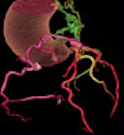
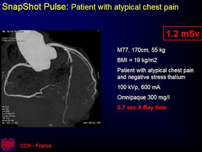
To reduce the CTA dose significantly, the team in St. Denis uses low-dose SnapShot Pulse acquisition, an axial step-and-shoot mode with prospective gating on a LightSpeed VCT XTG scanner (GE Healthcare, Chalfont St. Giles, U.K.).
The x-ray beam, triggered by ECG, is turned on only during the required heart phases and turned off during the rest of the cardiac cycle. Snapshot mode is used only when the heart rate is 65 bpm or lower to ensure that the best phase for coronary assessment is in diastole and when functional study is not required by the referring physician, Sablaoyrolles said.
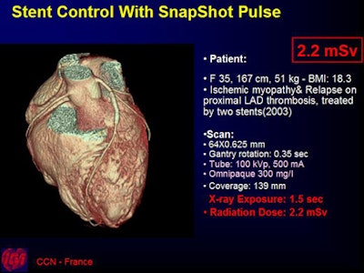
The acquisition parameters varied according to the body habitus of the patient and indications (105-280 coverage), ranging from 500-800 mAs and 80-120 kVp with slice thickness of 0.625 mm, 40-mm coverage/rotation, and 0.35-sec gantry speed. The group examined the image quality versus that obtained with the helical mode with retrospective gating.
After the administration of beta-blockers to reduce heart rates, SnapShot mode was used for patients with heart rates of 65 bpm or less (70% to 80% RR interval), and helical mode was used with ECG dose modulation for heart rates higher than 65 bpm (40% to 80% RR interval), Sablaoyrolles said.
Between September 2006 and August 2007, the team performed 1,155 cardiac exams in pulse mode (mean 5.6 mSv, DLP = 330, mGy.cm). An additional 276 aorta/bypass exams were done in pulse mode (mean 9.6 mSv, DLP = 560, mGy.cm).
The scan parameters are calculated "as a function of BMI of the patient," including the coverage, the mAs and kVp, and the padding of the x-ray window (70% to 80%).
The researchers did not detect any difference in image quality between the SnapShot Pulse exams and those performed in helical mode with retrospective gating.
 |
| All images courtesy of Dr. Jean-Louis Sablayrolles. |
For thinner patients, low-dose coronary CTA provides "equal or less radiation dose of a cath lab with the use of beta-blockers to control the heart rate," Sablayrolles said. Most cardiac exams are done this way, allowing radiation doses of 5 mSv or less using SnapShot Pulse. For these exams the iodine concentration can be lowered to, for example, 300 mg/L.
The new technique represents "a revolution in cardiac imaging, and a prerequisite for the future," he said.
Other researchers using SnapShot Pulse mode have reported mean radiation doses of 2 mSv or less for up to 97% of patients with heart rates 65 bpm or less, but Sablayrolles said his group decided to pad the x-ray window to reduce the chance of having to rescan a patient -- and by doing so forfeit part of the radiation dose savings.
 |
"Sometimes the best phase (for image reconstruction) is, for example, 80% not 70%, and I prefer a larger padding (of the x-ray window) to ensure we have the best phase," Sablayrolles told AuntMinnie.com. "Yet the dose reduction remains quite significant between prospective and retrospective gating," on the order of 60% to 70% dose savings, he said.
"This is very important: The image quality is the same" as retrospectively gated exams, he said. "Diagnosis of coronary artery disease is very difficult, so we need the best possible image quality." Still, the group did not report a systematic comparison of image quality results.
Low-dose coronary CTA might permit new indications for cardiac CT, for example, in asymptomatic patients with high risk factors, systematic bypass, or stent follow-up, he said.
By Eric Barnes
AuntMinnie.com staff writer
November 28, 2007
Related Reading
Radiation dose slashed in 64-slice coronary CTA, February 15, 2007
Step-and-shoot acquisition cuts CTA dose, improves images, January 16, 2006
Copyright © 2007 AuntMinnie.com


