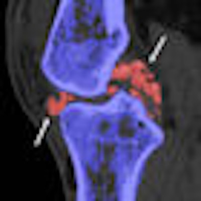
According to early assessments, dual-energy CT (DECT) has the potential to revolutionize the diagnosis of gout with a fast, sensitive, and noninvasive exam that outperforms the invasive gold standard test while revealing new information about an old nemesis.
Gout, the most common form of crystalline arthropathy and the most common inflammatory arthropathy in men, results from the precipitation of uric acid crystals within the joints.
The painful and destructive joint condition affects some 2.1 million people in the U.S. and the numbers are growing, said Dr. Savvas Nicolaou from Vancouver General Hospital and the University of British Columbia in Vancouver. The incidence of gout has been rising for a few decades, he said, spurring new interest in the disease. Where are the new cases coming from?
"This [increase] has coincided with high levels of soft drink and fructose consumption," said Nicolaou, who is director of emergency radiology at Vancouver General Hospital. "The high rate of fructose consumption results in increased uric acid. Two or more servings a day almost doubles an individual's risk of developing gout -- independent of other risk factors such as high alcohol consumption and high dairy consumption."
Of particular concern: an estimated 50% of the water consumption in the U.S. is being supplied through soft drinks, Nicolaou said.
The best known form of gout is an acute presentation in the tarsal joints of the foot, but the disease can actually affect any joint in the hand or foot, "and when left untreated may result in extensive joint destruction," Nicolaou said.
Gout is difficult to diagnose, too -- or at least it used to be. Certain clinical features can aid in diagnosis, but there is overlap with myriad conditions that can mimic gout, such as septic and rheumatoid arthritis, amyloid arthritis, osteoarthritis, sarcoidosis, xanthomatosis, and malignant intraosseous or para-articular masses (e.g., giant cell tumors), to name a few.
Definitive diagnosis of the monosodium urate crystal deposits requires polarized-light microscopic analysis of joint fluid aspiration, he said.
"Although joint aspiration is simple, it can be technically difficult, particularly in acutely inflamed joints, because there might not be any fluid to aspirate, and you might need US guidance to aspirate the fluid. There are also challenging anatomic sites such as the spine. A noninvasive means of diagnosis would be highly desirable," Nicolaou said.
In addition to the classic painful and visibly swollen joints, imaging can help find gout lesions. But radiography, single-source CT, MR, and ultrasound aren't specific enough to confirm a diagnosis, he said.
Radiographs are often normal-appearing until late in the diagnosis, by which time joint damage is already extensive. Single-source CT typically depicts gout as round or irregularly shaped opacities with attenuation in the range of 150-200 HU. Ultrasound yields limited sensitivity and specificity, depicting echogenic nodules with broad, indistinct acoustic shadowing that looks a lot like rheumatoid arthritis.
MRI offers variable depiction of gouty arthritis and tophaceous deposits, with intermediate intensity on T1-weighted imaging and intermediate-to-low intensities on T2-weighted imaging, Nicolaou explained. But even with MRI, "erosions in rheumatoid arthritis can have a similar appearance [to gout]," Nicolaou said.
But the prospect of fast, sure, noninvasive diagnosis with DECT (Somatom Definition, Siemens Healthcare, Malvern, PA) has spurred new interest in the disease at Nicolaou's institution, where he said Siemens' application was born.
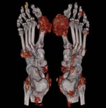 |
| At DECT, volume-rendered image of patient's feet shows extensive tophaceous deposits along metatarsal pharyngeal joints, with extensive tophi at midfoot and surrounding the ankle joint. Aspirationally proven gout deposits are depicted in red. All images courtesy of Dr. Savvas Nicolaou. |
Distinguishing uric acid
At Vancouver General, Nicolaou and his colleagues developed a dual-energy algorithm to identify uric acid stones, a method confirmed both in vivo and ex vivo to be highly sensitive for the detection of uric acid deposits. Dual-energy CT is used to tease out the differential attenuation values of uric acid deposits scanned at different energies, Nicolaou said.
"We worked on the renal uric acid protocol and modified it for gout," he told AuntMinnie.com. "Then Siemens used our data and scans to develop a new gout dual-energy protocol that anyone can now use." A commercial version of the application, Siemens' syngo DE Gout, received 510(k) clearance in March.
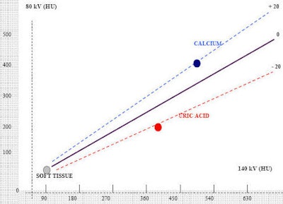 |
| Dual-energy CT software algorithm for gout detection relies on demonstrated lower CT values of uric acid deposits compared to calcium when the two materials are scanned at different energies (80 kV versus 140 kV). Red line indicates uric acid deposits; blue represents calcium. |
"We have an 80 kV dataset and a 140 kV dataset," he said. "They are loaded into a DECT viewer and 3D material decomposition algorithm is performed." For the dedicated gout protocol, the parameters have been manipulated to sensitize the software to uric acid in joints. Uric acid crystals representing gout are color coded red, with other bony formations represented in blue.
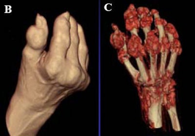 |
| Photograph of patient's hand (B) is shown with corresponding DECT image of aspirationally proven gout in red (C). Volume-rendered DECT image shows tophaceous deposits along the upper hand. |
The group performed a study to determine whether DECT could be used to diagnose gout, whether it was superior to clinical assessment, and further, if the modality could be used as a problem-solving tool in equivocal cases.
"We enrolled 37 patients with aspirationally proven gout," Nicolaou said. "They had chart review and also underwent a full dermatological assessment and DECT," which was read by two radiologists working independently.
DECT bests aspiration
According to the results, all patients with aspirationally proven gout had corresponding positive DECT scans. Conversely, DECT scans of a control group were all negative, he said.
"What we found is that DECT is far more efficacious than clinical assessment in identifying the presence of monosodium urate crystal deposition," Nicolaou said. DECT identified more gout sites per patient (19.5) than clinical assessment (4.61, p = 0.01).
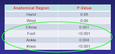
Subdividing the findings into anatomic regions, gout detection by DECT was significantly better than clinical assessment in the elbow, foot, ankle, and knee, but not the hand and wrist, he said.
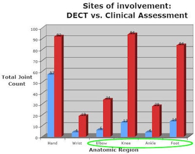 |
| When results from 37 patients are categorized into discrete anatomic regions, bar graph (above) shows significantly improved gout detection by DECT compared to clinical assessment in the elbow, knee, ankle, and foot. Conversely, DECT's advantage did not reach statistical significance (below) in the hand and wrist. |
 |
Perhaps more important, the study revealed new information about the progression and accumulation of monosodium urate crystal deposits.
"Surprisingly, the study revealed that uric acid deposits are predisposed to accumulate in tendons and ligaments," Nicolaou said. "This is very important because [the deposits] can predispose these ligaments and tendons to rupture. So if we can identify the disease early on, we can institute treatment and prevent destruction of tendons and ligaments."
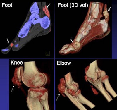 |
| New information about gout distribution: At DECT, crystalline uric acid buildup is detected in tendons and ligaments. |
DECT was also useful in detecting uric acid deposits in anatomic regions that are challenging to assess clinically and that represented subclinical disease, he said. Examples included intra-articular tophi within the knee and ankle joints, the collateral and cruciate ligaments of the knee, the popliteus tendons, and the deep flexor tendons of the wrist.
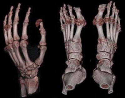 |
| Subclinical disease: Patient who had suffered a single gout attack a year earlier was found to have tophaceous gout at the tip of the thumb and first IP joint on the fourth digit at DECT. Further scans also showed uric acid deposits on the foot. |
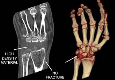 |
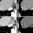
| Problem-solving: In a wrist trauma case, single-source CT (left) ruled out fracture. DECT (right) revealed high-density material to be uric acid deposits, proven at aspiration. |
"Up until now, no imaging modality had high sensitivity and specificity for detecting gout," Nicolaou said. "This novel technique provides a reliable and noninvasive means of diagnosing gout more sensitively than clinical examinations, confirming the burden of disease, distinguishing disease, monitoring response to treatment, and lastly, using it as a problem-solving tool."
By Eric Barnes
AuntMinnie.com staff writer
September 15, 2008
Related Reading
Low-energy, high-current CT nabs tiny liver lesions, May 15, 2008
Small lung nodules vex dual-energy DR, study says, May 1, 2008
Osteoarthritis may predispose to acute gouty attacks, October 15, 2007
Fluoroscopically guided steroid injection effective in hip osteoarthritis, August 22, 2007
Spiral CT protocol predicts kidney stone composition, May 19, 2003
Copyright © 2008 AuntMinnie.com

