A single dual-energy CT (DECT) exam can provide an accurate, integrative analysis of coronary artery morphology and myocardial blood supply, according to researchers from the Medical University of South Carolina in Charleston.
Dr. Balazs Ruzsics, Ph.D., and colleagues, who have been working on myocardial blood pool imaging for a few years now, believe the new test could save time, radiation, and money in the comprehensive evaluation of potential cardiac disease, primarily by eliminating the need for additional tests in patients with negative results.
"Preliminary evidence has shown that contrast-enhanced dual-source CT in dual-energy mode ('dual-energy CT') has the potential to evaluate changes in the status of the myocardial blood supply, in addition to the analysis of coronary artery morphology," they wrote in the American Journal of Cardiology (August 1, 2009, Vol. 104:3, pp. 318-326).
Because body tissues and intravascular iodine-based contrast media have unique spectral characteristics when exposed to different x-ray energy levels, DECT enables the mapping of iodine and, thus, blood distribution within the myocardium as a surrogate of myocardial perfusion and blood volume, they explained.
Importantly, this study of DECT in 36 patients obviated the need for further cardiac imaging exams in the 15 cases that were negative, highlighting the potential diagnostic efficacy of DECT as a one-stop shop. The results were well correlated to both invasive angiography and myocardial perfusion imaging.
"Using single-photon emission CT (SPECT), myocardial perfusion imaging, and invasive coronary angiography (ICA) as the reference standards, we evaluated the performance of DECT for integrative imaging of the coronary artery morphology and the myocardial blood supply," wrote Ruzsics and his colleagues, including Dr. Florian Schwarz and Dr. U. Joseph Schoepf.
The researchers retrospectively analyzed data from 36 consecutive patients (21 men; mean age, 57 ± 11 years) with equivocal or incongruous SPECT results who had undergone rest/stress SPECT myocardial perfusion imaging.
All patients had been referred for coronary CT angiography (CTA) for further evaluation of the SPECT results. Nine were sent to CTA for assessing graft or stent patency, 15 had positive SPECT results but a discordant clinical presentation, and 12 had persistent chest pain despite their negative SPECT results, the group reported.
Patients were examined on a dual-source CT system (Definition, Siemens Healthcare, Andover, MA) in dual-energy mode. Images were acquired with one of the x-ray tubes at 140 kV/114 mAs and the second tube set at 100 kV/165 mAs, using 0.6-mm collimation and a z-flying focal spot technique. No beta-blockers were used.
After administering an individualized contrast/saline injection and scout image acquisition, patients received a timed bolus of iodinated contrast (Ultravist, 370 mgI/mL of iopromide, Bayer HealthCare Pharmaceuticals, Wayne, NJ) followed by a saline chaser.
The DECT data were used to reconstruct anatomic coronary CT angiographic images and to map the myocardial iodine distribution within the left ventricular myocardium, Ruzsics and his team reported. Two readers working independently analyzed all the DECT studies for stenosis and myocardial iodine defects.
In addition, a segmental comparison was performed between the stress/rest SPECT perfusion defects and DECT iodine (perfusion) defects and between the invasive angiography and coronary CTA findings for stenosis.
Results
Compared to the SPECT results, DECT had 92% sensitivity and 93% specificity, overall, with 93% accuracy for detecting any type of myocardial perfusion defect, the group reported.
"Using SPECT, 17 patients had fixed myocardial perfusion defects in 89 segments and 13 had reversible myocardial ischemia in 68 segments," Ruzsics and colleagues reported. "Contrast defects on dual-energy CT correctly identified 85 (96%) of 89 myocardial segments with fixed and 60 (88%) of 68 with reversible perfusion defects.
On a per-patient basis, such contrast defects correctly identified 16 (94%) of 17 patients with fixed and 13 (100%) of 13 with reversible myocardial perfusion defects, they wrote.
Agreement between the readers was also strong (weighted k = 0.87), and the variability was nonsignificant (p > 0.05). Compared with angiography, coronary CT angiography had 90% sensitivity, 94% specificity, and 93% accuracy for the detection of greater than 50% stenosis.
"Diagnostic dilemmas arise when the findings from physiologic testing are equivocal or incongruous with the patient's overall clinical presentation," the group wrote.
DECT for myocardial imaging
Increasingly, the standard referral for invasive workup is replaced with coronary CTA to exclude coronary artery disease or refer the patient for additional invasive exams if findings are positive. In this study, the need for some of those additional exams was eliminated.
"Coronary CT angiographic findings suggestive of obstructive coronary artery disease resulted in the indication for ICA at our institution in 13 patients, and the exclusion of underlying significant coronary artery stenosis in 15 individuals obviated the need for additional cardiac evaluation of these patients," they wrote.
 |
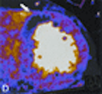
| Contrast-enhanced retrospective electrocardiogram-gated DECT in a 61-year-old man with atypical chest pain. Invasive coronary angiography (A) shows significant stenosis of the proximal left anterior descending artery by predominantly noncalcified plaque. Rest (B)/stress (C) SPECT myocardial perfusion imaging shows reversible ischemia in the anterior and anteroseptal myocardium (arrow). Corresponding DECT study at rest (D) delineates corresponding contrast defect (arrow). All images courtesy of Dr. Balazs Ruzsics, Ph.D. |
 |
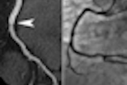
| Fifty-eight-year-old woman with recurring chest pain. Extensive, fixed perfusion defect is observed at stress (A) and rest (B) SPECT myocardial perfusion imaging. Contrast-enhanced retrospective electrocardiogram-gated DECT study: Coronary CTA reconstruction displayed as curved multiplanar reformat (C) shows total thrombotic occlusion of the distal left anterior descending artery to the first diagonal branch (arrow). Short-axis view (C) shows corresponding anteroseptal contrast defect (arrows) in good correlation with fixed perfusion defect at SPECT. |
Inserting coronary CTA into the diagnostic algorithm increases the cumulative radiation exposure, the authors wrote. However, for patients of the age group of this study in particular, "the stochastic risk of radiation-induced cancer is likely outweighed by the benefit of avoiding the small, but manifest, risks of invasive workup in patients without obstructive disease on [coronary] CTA."
The study builds upon the authors' previous work evaluating the myocardial blood volume using a non-time-resolved coronary CTA scan, they added.
"Because of this static nature, it would be imprecise to refer to our DECT technique as 'perfusion' imaging, because this would require time-resolved image acquisition of the passage of a contrast medium bolus through the myocardium," they wrote.
Rather, the technique exploits the material differentiation capabilities of cardiac dual-source CT in dual-energy mode to delineate myocardial areas with decreased contrast and, hence, blood, they content.
More research is needed on several fronts. For example, the observation that resting DECT can detect reversible perfusion defects that are visualized only during hyperemia on SPECT "is paradoxical in the context of the established paradigms of nuclear myocardial perfusion imaging," the authors wrote.
Nevertheless, other researchers have reported similar findings with single-source CT, "confirming our belief that our results are not spurious," Ruzsics and colleagues wrote. And compared with recent reports of similar techniques using single-source CT scanners, DECT has shown somewhat better performance in detecting SPECT perfusion defects.
"We hypothesize that DECT-based iodine mapping might be more sensitive for the detection of hypoperfused myocardium compared with hypoattenuation within the Hounsfield scale on which single-source CT is based," they wrote. "This hypothesis, however, needs to be confirmed by a direct comparison of single-source CT and DECT in the same patient, an approach that was naturally precluded by the retrospective nature of our analysis."
Further studies are also needed in a larger and less heterogeneous population, they added.
"Our initial experience suggests that DECT, as a single examination, might be promising for the integrative analysis of the coronary artery morphology and the myocardial blood supply and is in good agreement with ICA and SPECT," they wrote.
By Eric Barnes
AuntMinnie.com staff writer
August 10, 2009
Related Reading
Dual-source CTA turns in mixed results for coronary stenoses, July 30, 2009
Adenosine stress DECT equivalent to SPECT, MRI, November 21, 2008
Dual-source CT edges into cardiac SPECT turf, March 6, 2008
Study correlates CTA to angiography, myocardial perfusion SPECT, May 25, 2007
Stress MPI with Tc-99m SPECT identifies high-risk obese patients, September 1, 2006
Copyright © 2009 AuntMinnie.com


