Thursday, December 3 | 10:30 a.m.-10:40 a.m. | SSQ13-01 | Room N228
Perfusion CT offers additional functional information for the characterization of lymph nodes, which is useful for distinguishing benign from malignant nodes, according to a research team from India with extensive experience in perfusion imaging."Previous studies using CT, MR, and [ultrasound] have shown promising results in lymph node metastasis; however, they had low specificity and the lymph node size was the limiting factor," said Dr. Abhishek Mahajan from Tata Memorial Hospital in Mumbai.
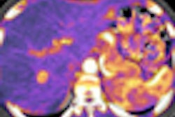
The group's prospective study examined 33 patients (age, 47-69 years) who were previously untreated and had head and neck, lung, and abdominal malignancies. Standard contrast-enhanced CT was followed by perfusion CT. The team compared perfusion characteristics, blood flow and volume, time to peak enhancement, mean transit time, and other measures, creating colored perfusion maps of the results.
The mean value of each parameter was calculated for every node separately, and the results were with the histological analysis of resected nodes, Mahajan said.
"On the perfusion maps, malignant nodes showed remarkable hyperperfusion compared to nonmalignant ones," he said. "The malignant [nodes] showed significantly higher blood flow values, blood volume values and peak enhancement values. Mean transit time was also much lower in malignant lymph node lesions. CT distinguished the malignant nodes with 91% accuracy.
"Information on vascularization provided by perfusion CT can be integrated with that of other diagnostic techniques to better understand changes occurring in lymph nodes," Mahajan said. "In doubtful cases it will be useful in differentiation between malignant and benign lymphadenopathy."


