High-pitch dual-source CT angiography (CTA) scans produce significantly less radiation-related DNA damage than comparable low-pitch dual-source CTA scans -- and, importantly, the DNA results correspond to both measured and derived biological radiation doses, say researchers from Erlangen, Germany.
Whether acquired with single- or dual-source CT, retrospectively gated 64-detector-row coronary CTA exposes patients to rather high radiation doses, with the advantage of enabling functional analysis of the heart. When functional analysis is not needed, however, significant dose reductions can be achieved using prospective tube current triggering in combination with dual-source CT.
The shortcoming with traditional dose measurements such as dose-length product (DLP), though, is that they don't address the interaction of the x-rays with the patient's body, said Dr. Michael Küfner from the University of Erlangen.
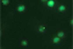
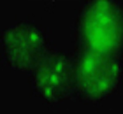
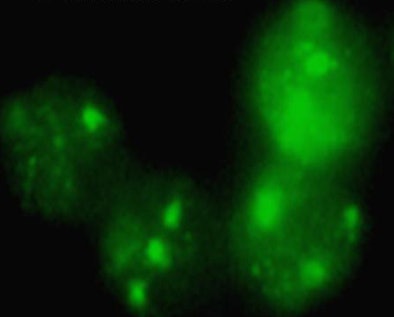
"Previous biological dosing approaches failed due to low sensitivity of the techniques, but the determination of DNA double-strand breaks [DSBs] using a novel immunofluorescence microscopic method provides an accurate estimate of biological radiation effects," he said in a presentation at the 2010 European Congress of Radiology (ECR). "The histone variant H2X is phosphorylated when exposed to radiation, and the resulting γH2AX can be visualized using a fluorescence microscope. We can see tiny green spots within the nuclei, and each of the foci represents one double-strand break."
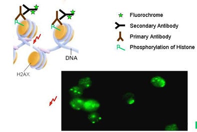 |
| Double-strand breaks are among the most significant DNA lesions introduced by ionizing radiation. The lesions can be visualized indirectly using foci of phosphorylated histones as a surrogate for DSBs, via γH2AX immunofluorescence microscopy. Small green foci in image below represent DSBs. All images courtesy of Dr. Michael Küfner. |
 |
The researchers examined blood samples obtained from 34 patients (ages 26-82 years; median, 57.5 years) undergoing coronary dual-source CTA using either 64-detector-row dual-source CT (Somatom Definition, Siemens Healthcare, Erlangen, Germany) or 128-detector-row dual-source CT (Somatom Definition Flash, Siemens).
Acquisition parameters for each scanner were as follows:
- 128-detector-row (n = 15): high-pitch spiral scan, 38.4-mm collimation, 100-120 kV, 320-456 mAs/rotation, 3.2-3.4 pitch
- 64-detector-row (n = 19): low-pitch spiral scan, 19.2-mm collimation, 100-120 kV, 330-438 mAs/rotation, 0.2-0.39 pitch, electrocardiographic pulsing
"We collected blood samples from all of the patients before and after the CT scan, isolated the blood lymphocytes, and determined the DNA DSB using the immunofluorescence microscopy technique," Küfner said.
The actual number of double-strand breaks was calculated by subtracting the baseline DSB levels obtained before exposure to the number of scans. The group also calculated a biological dose measurement by performing an in vitro radiation exposure of each patient's blood at 50 mGy. Then, by comparing the values obtained in vivo with those obtained in vitro, the researchers were able to assess the biological dose, which they called radiation dose to the blood.
The number of double-strand breaks resulting from radiation in vivo was calculated by comparing prescan DSB levels to those obtained immediately thereafter.
According to the results, total dose-length product ranged from 101 to 237 mGy/cm in the high-pitch scans and from 524 to 1,283 mGy/cm in the low-pitch scans (p < 0.0001). DSB levels obtained 30 minutes after the CT scans ranged from 0.02 to 0.7 per lymphocyte.
"There was a strong correlation between the double-strand breaks and the DLP, and patients undergoing the high-pitch protocol had both lower DSB levels and lower DLP," Küfner said.
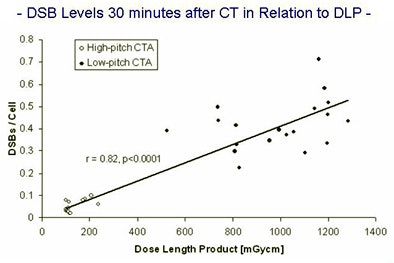 |
| The number of DSBs shows good in vivo correlation to DLP (r = 0.82, p < 0.0001) 30 minutes after high- or low-pitch CTA scans. High-pitch scanning lowered the dose by 70% to 90% overall. |
In addition, DSB levels 30 minutes after exposure were significantly lower after high-pitch scans (median, 0.04 DSBs/cell; range, 0.02-0.10) compared to 30 minutes after the low-pitch scans (median, 0.39 DSBs/cell; range, 0.22-0.71, p < 0.0001).
Finally, the researchers found good correlation between the radiation dose to the blood and the dose-length product (r = 0.82, p < 0.0001).
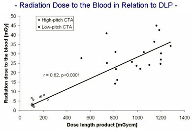 |
| Dose-length product shows good in vivo correlation to radiation dose to the blood (r = 0.82, p < 0.0001) following high- or low-pitch CTA scans. |
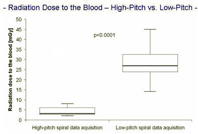
The radiation dose to the blood was significantly reduced in the high-pitch group (median, 3.1 mGy; range, 2.0-8.1) compared to the low-pitch group (median, 26.9 mGy; range, 14.2-44.9, p < 0.0001).
"We have a 90% reduction of the dose-length product by using the high-pitch spiral data acquisition compared to the low-pitch protocols, and we also find a 90% reduction of the biological dose," Küfner said. "By using the Flash [high-pitch] scans these differences were statistically significant."
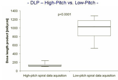 |
| High-pitch dual-source scans delivered significantly less radiation than low-pitch dual-source scans as measured by dose-length product (above) as well as radiation dose to the blood (below). |
 |
One inconsistency in the scan protocols merited further analysis, Küfner said. Most patients in the high-pitch group were scanned at 100 kV, compared with 120 kV in the low-pitch group -- a factor that biased the results in favor of the high-pitch scans.
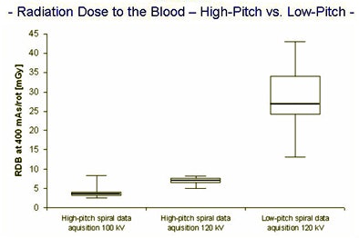 |
| Dose to the blood results for high-pitch versus low-pitch spiral acquisitions were adjusted to reflect overall higher kVp values in the low-pitch scans, which initially biased the results in favor of lower doses for high-pitch scans. After compensating for the bias, high-pitch scans still yielded dose savings of approximately 70% versus low-dose spiral acquisitions. |
After adjusting the results to compensate for the kV level disparity, however, patients scanned with the high-pitch protocol still received a dose that was about 70% (rather than 90% in the unadjusted calculation) lower than the low-pitch scans.
CT scanning produced DSBs in all patients, and "using our immunofluorescence microscopy technique, we found a reduction in DNA double-strand breaks in all of our patients," Küfner concluded.
"Thirty minutes after coronary CTA, we found rather high DNA damage levels, especially in low-pitch patients," he said. "The median was 0.39 DSB per cell. That means that in every second or third lymphocyte, the double-strand break is induced by the CT scan."
By Eric Barnes
AuntMinnie.com staff writer
April 9, 2010
Related Reading
FDA hearings rise above medical radiation rhetoric, March 31, 2010
Firm promotes BioShield pill to address dose dilemma, September 15, 2009
Biological dose measures promise new view of cardiac imaging risk, May 1, 2009
Bisphosphonates may prevent radiation-induced leukemia, April 21, 2009
Researchers find genes that affect radiation damage, April 9, 2009
Copyright © 2010 AuntMinnie.com


