SAN FRANCISCO - Backed by growing clinical evidence, radiologists should step up to the plate and cut the tube voltage in their cardiovascular CT protocols. But getting the benefits without the pitfalls means paying attention to some technical considerations, according to a presentation at this week's International Society for Computed Tomography (ISCT) meeting.
Reduced kVp is associated with a reduction in radiation exposure without loss of image quality when used appropriately, said Sandra Halliburton, Ph.D., from the Cleveland Clinic Foundation in Ohio. And you get to cut the contrast dose, too, for a safer exam, she said. But there are few official guidelines, so a little improvisation will be in order.
"You can get some really nice low tube voltage images, but 120 kVp still seems to be some kind of standard," Halliburton said in her talk.
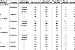
In the absence of real standards, kVp levels are usually based on visual assessment of patient size. Sometimes more objective measures such as patient weight and body mass index are used. Several studies, mostly from Europe, have suggested 100 kVp for patients weighing 85 kg or less. One group recommends 80 kVp for patients weighting 60 kg or less.
The studies represent a kind of standard that radiologists can look to in the absence of true guidelines, Halliburton said.
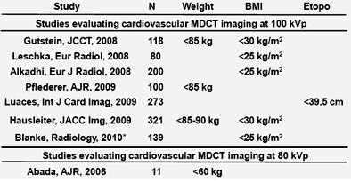 |
| Studies support the use of low-kVp cardiovascular imaging. All images courtesy of Sandra Halliburton, Ph.D. |
Radiologists selecting kVp should also consider the tube voltage capacity, she said. "In practice, many people have been disappointed with lower tube voltage imaging because of excessive noise in the image, but some of that can be compensated for, to the extent that our tubes allow, with an increase in tube current," she said.
Less contrast, more attenuation
Lower tube voltages increase iodine attenuation significantly, a fortunate circumstance that offers an opportunity to reduce the iodine concentration, Halliburton said.
Reduced kVp has "practical implications with the use of bolus monitoring, which is common in many applications," she said. With bolus monitoring, "when a certain attenuation threshold is reached, data acquisition is triggered. If we make no changes to our contrast protocol, we're going to see a shift upward of the attenuation curve, and subsequently, perhaps, an early triggering of our scan. That's something a lot of people don't think about when they choose to change the tube voltage."
The attenuation threshold for triggering the acquisition can be raised to compensate, of course, but the real solution is to lower the contrast dose by using a lower-concentration contrast agent, diluting contrast with saline, or reducing the contrast agent flow rate, she said.
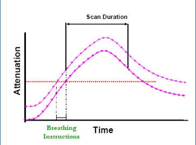 |
| Lower kVp raises the attenuation of blood, which can cause bolus timing to trigger an acquisition too early. But rather than raise the threshold for triggering, it's better to reduce contrast dose. |
"In some patients, particularly smaller ones, failure to modify contrast injection with a lower tube voltage can actually result in luminal and blood pool attenuation that obscures areas of interest, particularly if you're trying to delineate, say, coronary calcium," Halliburton said.
Quantitative image analysis
Also bear in mind that tissue attenuation characteristics change with photon energy -- and that the change is greater for higher-attenuating tissues, Halliburton said. As a result, HU-dependent analysis is impacted by changes in tube voltage, which affects the following:
- Differentiation of simple fluid and blood products or soft tissue
- Quantification of coronary calcium
- Characterization of coronary artery plaque
- Detection of myocardial infarction
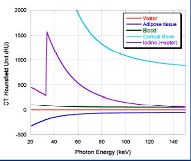 |
| Tissue attenuation characteristics fluctuate with changes in photon energy, and the changes are greater for higher-attenuating tissues. |
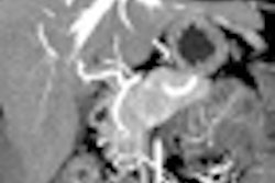
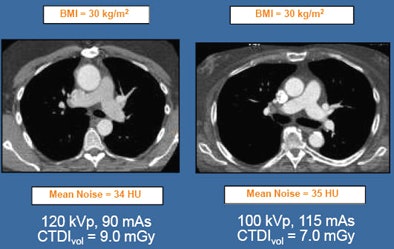
Because radiologists rely on attenuation to differentiate simple fluids, be aware that their attenuation can change slightly with reduced kVp imaging, she said. For example, a thrombus that measured 34 HU at 140 kVp measured 36 HU at 80 kVp -- a minor difference that might not be noticed. With a high-attenuating material such as calcium, however, the effect is more pronounced. In one case, for example, the attenuation of coronary artery calcium (below) rose from 130 HU at 120 kVp to 147 HU at 100 kVp.
 |
In 2009, Maurovich-Horvat et al evaluated the dimensions and attenuation of coronary artery plaque types (lipid-rich, fibrous, calcified) in a dozen coronary CT angiography studies in an effort to "improve our understanding of the natural history of coronary artery disease as well as prevention of cardiovascular events," they wrote (Journal of Cardiovascular Computed Tomography, November-December 2009, Supplement 2:S91-98).
Another study found that increasing the attenuation threshold could deliver comparable Agatston and calcium volume scores at different tube voltages, Halliburton said.
Most of the attenuation values in the literature are based on 120 kVp, so an appreciation of emerging reference levels for low-kVp imaging is important for success, Halliburton said. CT also relies on HU values to identify perfusion defects in the myocardium; these, too, will change with low kV imaging.
Standards are needed for the selection of tube voltage and current based on objective measure of patient sizes, Halliburton said in summary.
"Selection of a lower tube voltage should be balanced by an increase in tube current to the extent possible to maintain image noise," she said. "Lowering the tube voltage may not be appropriate when HU-dependent analysis is required."
Finally, contrast administration should be modified for lower tube voltages to ensure proper contrast timing, avoid excessive blood pool attenuation, and reduce contrast load, Halliburton said.
By Eric Barnes
AuntMinnie.com staff writer
May 19, 2010
Related Reading
Lower-kilovoltage coronary CTA maintains image quality, August 5, 2009
Peak voltage cut improves pulmonary CT angiography, July 22, 2009
Tube current modulation cuts triple rule-out CTA dose, April 15, 2009
Low-energy, high-current CT nabs tiny liver lesions, May 15, 2008
Dual-source CT edges into cardiac SPECT turf, March 6, 2008
Copyright © 2010 AuntMinnie.com


