CT in 2010 is lightning-fast, technically dazzling, and more popular than Miley Cyrus at a tween slumber party. It's invaluable for trauma and stroke patients presenting to emergency departments, and for ruling out worry in patients with chest pain.
At this year's RSNA conference, technical innovation leading to speed and image sharpness continues to make the modality popular with radiologists and bean counters alike.

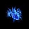
Functional imaging of the brain and heart are going low-dose and high-output. Mayo Clinic researchers, for example, are experimenting with brain perfusion images acquired at the dose of a normal head scan. In the heart, one group is finding success with dynamic myocardial stress CT perfusion, getting favorable results versus MRI. In another experiment, investigators are hoping to replace an expensive biomarker test with CT scans that were being done anyway.
Meanwhile, tumor perfusion imaging is being applied to different kinds of cancers -- for example, treatment monitoring in soft-tissue sarcomas, or malignant lung nodule detection by Japanese researchers. And of course, the use of dual-energy CT to separate scanned materials, such as renal stone types, is becoming increasingly sophisticated. Computer-aided detection is growing faster and delivering fewer false positives, while other researchers are using CT to quantify emphysema and air trapping.
For juggling the complex world of multimodality digital media and design, we recommend an RSNA/American Association of Physicists in Medicine (AAPM) plenary session, PS50, titled "Slicing Through Complexity in Medical Imaging," being held on Thursday, December 2, from 1:50 p.m. to 2:45 p.m. in the Arie Crown Theater.
Below you'll find a smattering of some of the innovative CT scientific presentations you might consider attending. You'll definitely want to visit the RSNA meeting program website as well to round out your conference schedule. Have a great meeting!