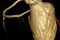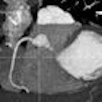
There are significant differences in the way 320-detector-row and 64-detector-row CT scanners perform heart scans of chest pain patients in the emergency room, according to a study presented at the recent Society of Cardiovascular Computed Tomography (SCCT) meeting in Denver.
While the authors found that radiation doses were similar, the 320-detector-row scanner used less contrast and had fewer artifacts and fewer nondiagnostic studies, even though the 64-detector-row images appeared a little sharper.
Cardiac CT is increasingly used to scan chest pain patients in the emergency room; however, few studies have compared the performance of different scanner models, particularly 320-detector-row CT (Aquilion One, Toshiba Medical Systems) to 64-detector-row CT (LightSpeed VCT, GE Healthcare).
The study, performed at Stony Brook University Medical Center in New York, retrospectively looked at the use of two scanners in the emergency department in 2009 and 2010, Dr. Melissa Daubert told AuntMinnie.com in an interview.
"The limitation is that we didn't have a similar protocol between two tests," because both scanners were used for cardiac CT scans as well as the longer triple-rule-out protocols that cover the upper chest, she said. There were other minor differences in the protocols as well. For example, the 64-detector-row scans required a test bolus of contrast media, while the 320-detector-row scans used automatic bolus tracking.
In all, the group scanned 449 patients using 320-detector-row CT and 261 with 64-detector-row CT. Of these, 308 were triple-rule-out scans, Daubert said. A total of 402 patients were scanned with the cardiac-only protocol, including 210 who underwent 64-detector-row CT versus 192 patients at 320-detector-row CT during the study period.
The study included patients with chest pain and low to intermediate probability of coronary artery disease. Exclusion criteria included nonsinus rhythm, contraindication to contrast media, and use of beta-blockers.
The results showed that many more patients in the 320-detector-row group were able to be scanned with prospective electrocardiogram gating, which tends to lower the radiation dose: 91.7% of the 320- versus only 77.1% of the 64-detector-row scans (p < 0.001), Daubert and her colleagues reported.
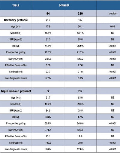 |
| Coronary CT angiography and triple-rule-out results, 320- versus 64-detector-row CT. Table courtesy of Dr. Melissa Daubert. |
"The 64-row scanner takes five to six revolutions [to cover the heart], so if you don't have a regular heart rate for at least six to seven beats, it will go to retrospective gating to make sure you get a diagnostic study," she said. The 320-detector-row CT has a larger 16-cm anatomic coverage per revolution, "so all you need is one really good beat" to cover the heart in cardiac mode -- two beats for triple-rule-out scans.
Also, probably due to the smaller number of beats required to capture the heart at 320-detector-row CT, "we actually found a statistically significant difference [p < 0.001] in the percentage of nondiagnostic scans: 2.6% at 320, while the 64 had 5.7%," Daubert said.
Other findings included the following:
- The 320-detector-row scans required significantly less contrast (71.0 ± 0.6 cc versus 97.7 ± 0.8 cc, p < 0.001).
- However, there was no significant difference in the mean effective radiation dose (7.56 ± 0.34 mSv [320] versus 6.58 ± 0.43 mSv [64].
- Similar results were seen in patients undergoing the triple-rule-out protocol, although this protocol showed a trend toward reduced effective dose at 320-detector-row CT (9.51 ± 0.39 mSv [320] versus 13.12 ± 1.91 mSv [64], p = 0.069). The difference in contrast requirements was also more pronounced.
In theory, the 320-detector-row machine should produce significantly lower effective dose; however, in this case, the use of more than one kV setting may have confounded the dose results, according to Daubert. It was the scanner software that chose 80 kV or 100 kV, and the 64-detector-row machine favored 80 kV more often than the 320-detector-row CT for reasons unknown.
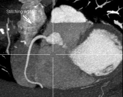 |
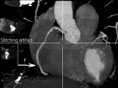
| Owing to the need to acquire cardiac images over several heartbeats, coronary CT angiography at 64-detector-row CT produces more artifacts than 320-detector-row CT, which acquires the entire cardiac volume in a single beat, or two beats for a longer triple-rule-out study. Above, 320 image acquired over two heartbeats has a single stitching artifact (arrow), while the 64 image below contains four important artifacts, only one of which is labeled with an arrow. Can you find the rest? Images courtesy of Dr. Melissa Daubert and Dr. Michael Poon. |
 |
"The 64-slice machines were able to image a lot more patients at the lower 80-kV voltage," she said. "We'd like to look at it in randomized fashion" in a prospective study that compares equivalent kV settings, "because it's suggested that when you use the same protocol, 320-slice will have lower radiation."
The main advantage of 320-detector-row CT is the full volumetric scanning that delivers a sharp reduction in artifacts due to scanning the heart in a single beat, she said. As a result, it produces fewer nondiagnostic studies versus 64-detector-row, which acquires the volume of the heart over several beats.
If the heart rate is low enough, "sometimes they both come out beautifully and you can't tell the difference between the scans," she added, but even in diagnostic studies small artifacts are often visible at 64-detector-row CT.
On the other hand, "the 64 tends to give smoother images," she said. "The 320 seems to be a little grainier even when you control for the size of the patient -- I don't know why that is." A more complete analysis of image quality differences is forthcoming, Daubert added.
Overall, the study found that 320-detector-row coronary CT angiography was associated with lower contrast use and equivalent radiation exposure versus 64-detector-row CT, the group concluded.


