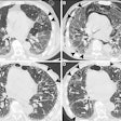
Lung cancers identified in low-dose CT screening programs are similar to those identified outside of the screening setting, according to results released on March 27 in Radiology. The study may allay concerns that cancers detected by screening are less aggressive and therefore warrant less attention and resources than cancers identified by other means.
The study is based on data from the International Early Lung Cancer Action Program (I-ELCAP), a multicenter clinical study in which patients were screened annually with CT between 2003 and 2009 using a common protocol. Researchers found that the mean volume doubling time of screen-detected cancers was about 100 days, similar to cancers diagnosed as a result of symptoms.
Moreover, the frequency of small-cell carcinoma and adenocarcinoma were similar for cancers detected in screening versus cancers detected outside of the screening program (Radiology, March 27, 2012).
"There's always been discussion of what type of cancers does screening pick up -- some people have said they're all so slow growing that they wouldn't have killed you -- so what we did is we looked at cancers you detect in annual rounds which should reflect ... what you find in usual care," said lead author Dr. Claudia Henschke, in an interview with AuntMinnie.com. "We compared it to the data that has been published extensively about cancer found in clinical care, and we found that [screened cancers] were very like what you find in clinical care."
"These are real cancers that would kill you if they weren't discovered early, so it kind of underscores again the data that we had shown in ELCAP and also that NLST [National Lung Screening Trial] has shown -- that screening for lung cancer saves lives." Henschke is a professor of radiology at Mount Sinai Medical Center in New York City.
The investigators also found that subsolid lesions grew more slowly than solid lesions, potentially enabling less frequent follow-up for subsolid lesions.
Cancers diagnosed in repeat screening rounds should be similar to lung cancers diagnosed outside of screening programs; however, this question had not been well-examined, the team wrote. The matter is also important because if cancers detected in repeat screening are less aggressive than those detected in other settings, the resources spent for repeated screening rounds of these patients may be wasted, and the radiation doses may be unnecessary.
Volume doubling time
The study aimed to empirically address the distribution of the volume doubling time (VDT) of lung cancers diagnosed in repeat annual rounds of CT screening in the I-ELCAP registry, with regard to tumor growth rates as well as cell types.
The study team identified all instances of first primary lung cancer diagnosis after a negative screening result between seven and 18 months earlier, including repeat screenings undertaken when symptoms appeared between screenings. Lesion diameter was calculated by measuring the length and width of each cancer at the time the nodule was first identified for further workup, and after the most recent prior screening. Using these two time points, the length and width were measured a second time for each cancer, and the results were used to calculate VDT, Henschke and colleagues explained. VDT distributions were compared using the chi-square test statistic.
Of the 111 cases, 110 (99%) were diagnosed at screening and one (1%) was prompted by symptoms between screenings; 88 (79%) were stage I. Fifty percent of the cancers were adenocarcinomas, 19% were squamous-cell carcinomas, and 13% were cancers of other cell types, the authors reported.
In nonscreening settings, the frequencies of small-cell carcinoma and adenocarcinoma among all lung cancers have been reported to be approximately 20% and 50%, respectively. Thus, the study results from repeat rounds of CT screening, 19% and 50%, respectively, were nearly identical, the authors noted. In addition:
- The median VDT for all 111 cancers was 98 days (mean, 136 days). Breaking down lesions by VDT, 56% of the cancers doubled in less than 100 days, 31% doubled in 100-199 days, 13% doubled in 200-299 days, 4% doubled in 300-399 days, and 3% saw doubling times of 400 days or more. For the 90 non-small cell cancers, the median VDT was 121 days (mean, 154).
- Small-cell cancers were the fastest-growing lesions (median, 43 days), followed by large-cell/neuroendocrine cancers (median, 82 days), squamous cell cancers (median, 88 days), adenocarcinomas manifesting as solid nodules (median, 140 days), and, finally, adenocarcinomas manifesting as subsolid nodules (median, 251 days).
- Solid nodules represented 89% (n = 99) of the 111 cancers, while the 12 subsolid nodules were all adenocarcinomas that grew much more slowly. The distribution of the VDTs for cancers manifesting as solid nodules and that for cancers manifesting as subsolid nodules was significantly different (p < 0.001).
"Relevant to the VDTs reported here, it is of note that adenocarcinomas manifesting as subsolid nodules do not only progress by increasing in size but also by internal growth that is manifested as increasing CT attenuation and/or the development of solid components without an increase in external diameter," Henschke et al wrote. "Therefore, our measured values for the VDTs of these cancers may well be quite conservative."
Distribution of lung cancers diagnosed in repeat annual screening
|
Subsolid nodules were significantly less aggressive than solid nodules (p < 0.001).
The VDTs were very similar to results obtained in a nonscreening setting. A recent study of nonscreen-detected lung cancers by Detterbeck and Gibson, one of the only papers to differentiate between screen-detected and nonscreen-detected tumors, found a median VDT for non-small cell carcinomas of 121 days (mean, 135 days) with values of less than 100 days for 44% of tumors and more than 400 days for 4%. The numbers are very similar to the results of Henschke et al.
While noting as a study limitation that the group's use of a method for determining lesion diameter by length and width assumes sphericity and may not adequately reflect the complexity of the lesion, the method has been widely used in nonscreening studies of lung cancer, the authors stated.
Of course, volume doubling time isn't the answer to every question about lung cancer screening with CT, particularly with regard to subsolid nodules, and more research is needed to continue improving physicians' understanding of these issues, Henschke said.
"The doubling time is not everything, and in fact there's another paper that appeared in this month's Radiology [April 2012, Vol. 263:1, pp. 279-286] that showed how you can get an idea of the internal growth," she said. "You have to get an idea of the internal growth of these subsolid lesions as well as the change in size, so what we're saying is that we still need to do further work and research, but that perhaps the diagnostic work and the treatments can be tailored differently for subsolid lesions than they are for solid lesions."
For example, research in ground-glass opacities is progressing, Henschke said. When these lesions persist over time and increase in density -- not necessarily in size -- "then you really have to consider cancer as a possibility," she said. "All of the ones that we found in those kind of nodules have been adenocarcinomas."
Overdiagnosis not a problem?
The study results demonstrate that overdiagnosis is not the problem in lung cancer screening that some feared it would be, and that screen-detected cancers are as aggressive as those detected in other settings, the authors concluded.
"The comparability of the VDTs for lung cancers diagnosed in annual rounds of CT screening with those of cancers diagnosed in clinical practice tends to refute the two assertions that have been made regarding growth rates of the lung cancers diagnosed at CT screening," they wrote.
"One is that the screening tends to identify cancers that are so slowly growing that they would not become life threatening -- that is, that the screening results in major overdiagnosis. The second contention is that the life-threatening cancers grow so fast that they cannot be detected at screening while still curable."
The evidence behind these assertions has largely relied on the interpretation of chest radiography screening trial results "in which no mortality benefit was demonstrated but an excess of early-stage lung cancers was identified in the screening arm," they wrote. "These ideas have led to viewing screen-detected lung cancer as potentially representing a disease entity of its own, one that is not a precursor of advanced-stage disease."
The NLST clearly showed that screening saves lives using the traditional approach of which nodules to follow up and when, Henschke told AuntMinnie.com. But although NLST showed a 20% mortality reduction, it wasn't designed to show the maximum mortality reduction, which is probably much higher than 20%, she said. "So if you think about some 50-year-olds and other people who are at more risk because they smoke more by that age," you might consider using the National Comprehensive Cancer Network (NCCN) screening guidelines which extend the entry criteria to other individuals at high risk.
"So I think there's certainly more research to be done as to what ages screening should start at and what the criteria for risk are," she said, and the maximum mortality benefit also comes from continued repeat screening rounds -- more than just the three rounds included in NLST.
As for future research, "we continue to do research on better integrating smoking cessation into a screening program and coming up with optimum approaches," Henschke said. A new Veterans Affairs study in Phoenix is doing just that, she added. Another key area of research is in people who have never smoked, a population that is now responsible for about 30,000 lung cancer deaths per year.
"We really think that as screening practice develops, ideally, you would continue to accumulate the information on people who were screened, so that you continue to optimize the screening program as technology advances," Henschke said. "The CT scanners we have now are really phenomenal," with resolution that continues to improve as the radiation dose falls, "so that the amount of information you can get out of them for emphysema, for coronary artery risks, and so on, continues to increase."
