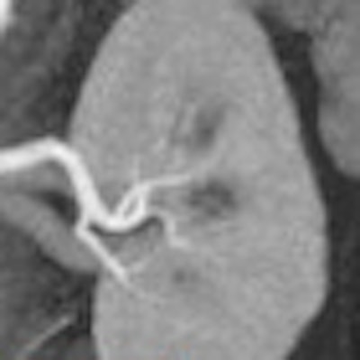
CT angiography (CTA) of the abdomen is a well-established method for answering any number of diagnostic questions, but in the past couple of years it's gotten even better. Recent technical advances, such as spectral imaging CT at different energy levels, can improve the modality's diagnostic power, reducing dose and contrast use.
That's according to a presentation from Dr. Dushyant Sahani from Massachusetts General Hospital, who spoke in June at the International Society for Computed Tomography (ISCT) annual meeting in San Francisco. Sahani discussed several emerging improvements in hardware, software, and imaging techniques for abdominal CTA that make the radiologist's job easier and the patient safer. In the new paradigm, lower contrast doses go hand in hand with reduced radiation exposure, he said.
"In the past, we assumed that our contrast agents were safe for everyone, but increasingly we recognize that there are certain individuals at risk, and we need to make our technologies and contrast use safer for these patients," Sahani said.
Interest is also growing in imaging-based biomarkers that help stratify patient risk and monitor response to various new treatments, he said. Most important are developments in the intravascular signal, particularly in low-kVp imaging, which is a powerful way to get more for less when used in patients with appropriate body weight. In the newest applications, the process is even automated, he said.
Low-kV imaging is among the best known and most effective new ways to cut dose and contrast at the same time, according to Sahani. Reducing tube peak energy (80-100 kV) leads to an increase in image contrast, an effect that occurs because the energy in the x-ray beam moves closer to the k-absorption edge of iodine.
If you are acquiring the low-kV image with dual-energy CT, there are two big benefits, he said. First, you can use the higher-energy data to look at organs and the low-energy data, which increases iodine enhancement, to look at vascular structures.
"In the past, there was some concern about image quality in the low-energy dataset; however, these concerns have been overcome by better use of energies based on patient size and better use of image settings at the two different energies," Sahani said.
"One can also use a combined or optimal-contrast image, which is a combination of the low- and high-energy image, to look at vessels if the low-energy image is noisier in certain subjects," he said.
Optimal-contrast images use voxel-based weighting of dual-source acquisitions based on native CT numbers, offering less noise and more contrast than linear average images.
Detector technology
Advances in detector technology, such as the Gemstone technology from GE Healthcare, are also improving the picture for abdominal imaging by providing high-resolution dual-energy imaging on a single-source scanner.
The technology "can enable processing of the dual-energy images to create virtual monochromatic images from 40 to 140 keV, and there are clear signs that using virtual monochromatic images for improving vascular enhancement is a real benefit because it gives you an additional series of monochromatic images to exploit for better diagnostic performance," Sahani said.
Virtual monochromatic images are synthesized from material density images. They depict how the imaged object would look if the x-ray produced photons at a single energy.
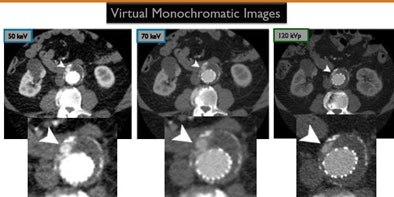
Examining monochromatic images acquired at 40 kV to 140 kV, for example, shows that at 50 kV there is more than twice the Hounsfield attenuation within the intravascular structures compared to standard polychromatic images, Sahani said. At 70 kV, the contrast drops, but image quality improves slightly and is still better than conventional 120-kVp images. In the case of a subtle endoleak, for example, one will be able to diagnose it more confidently in lower-keV images, and there are other techniques such as decreasing contrast volume to improve vascular enhancement.
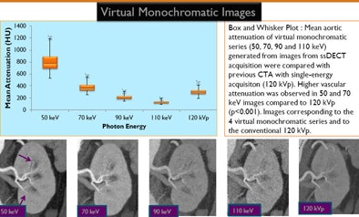 |
| Above, Box and Whisker plot shows mean aortic attenuation of virtual monochromatic series (50, 70, 90, and 110 keV) generated from single-source, dual-energy CT compared with previous CTA using single-energy acquisition (120 kVp). Below, higher vascular attenuation was observed in 50- and 70-keV images compared to 120 kVp (p < 0.001). Images correspond to the four virtual monochromatic series. All images courtesy of Dr. Dushyant Sahani. |
 |
Photon-counting detectors
Several CT vendors are also working on photon-counting detectors, which use a single x-ray tube and detectors that can discriminate high from low energy, Sahani said. Each x-ray photon absorbed in the detector creates an electronic pulse, and the height of the pulse is proportional to the energy of the absorbed x-ray photon. A pulse height discriminator sorts the pulses into two groups, high and low, and from these data the CT image is reconstructed.
With the photon-counting detector discriminating the energy level of each photon, the polychromatic beam is reprocessed to create virtual monochromatic images. The result is both lower dose and the benefits of multienergy data acquisition that improves vascular images.
Postprocessing techniques
The new imaging modes do create workflow challenges, however. The large number of images acquired in a single vascular CTA scan means that workflow demands automation.
"It's clear that we cannot analyze these on a conventional basis, looking at the raw data and the number of axial datasets," Sahani said. Automated postprocessing techniques will make workflow manageable, and the use of these techniques will facilitate rapid and efficient imaging evaluation that is less user-dependent.
Advanced vessel analysis encompasses a wide range of applications that facilitate examining vessels, such as automated centerline detection of multiple vessels, curved or stretched maximum intensity projections (MIPs), automated measurement of cross-sectional quantities along the vessel path, and plaque and bone subtraction tools. These are especially useful in small and calcified vessels and in vessels with stents.
"One of the benefits of CTA is higher contrast," he said. "A lot of the software can be automated because it can recognize the iodine intravascular attenuation and can segment the vessel, and assess the site of the vessel and the patency of structures."
Strategies to facilitate stent patency analysis include acquisition with thinner collimation, dual-energy CT with virtual monochromatic images, metal artifact reduction software (MARS) to reduce motion artifact, high-spatial-resolution reconstruction kernels, and vessel segmentation in postprocessing.
The tools are useful in presurgery planning as well as diagnosis.
"We can apply advanced tools in their desired plane but also segment various other organs for the benefit of our surgical colleagues in critical decision-making for major surgeries in the abdomen," Sahani said.
"Assessing stent patency can be very challenging ... especially in the smaller caliber vessels" such as the superior mesenteric and renal arteries, he said. With stent evaluation, radiologists are looking for restenosis attributable to intimal hyperplasia. But blooming artifacts caused by the stents may impair quantification of stenosis and create artificial lumen narrowing.
In the past, problems visualizing these small structures have been addressed by acquiring high-resolution images at a very low pitch and a high radiation dose, he said. But now radiologists can exploit dual-energy imaging to separate the calcium and iodine from metal, enabling the reliable visualization of intravascular structures to assess their patency.
In addition, there are algorithms to suppress artifacts, allowing more confident assessment of vascular patency. The technique is fast, and it doesn't introduce misregistration artifacts, he said.
Patient safety bar keeps rising
Patient safety is an increasingly important issue that relies on reducing the contrast as well as radiation dose, Sahani said. Using less contrast is critical for patients with impaired renal function. With low-kVp imaging, it's possible to drop the contrast dose way down.
"For older patients who are at risk for renal insufficiency, we often use just 40 to 60 mL of iodinated contrast media and improve the intravascular enhancement by either 80- or 100-kVp scanning or employ a dual-energy technique," he said. Obviously, reduced dose of iodinated contrast material also provides an ancillary benefit of some cost savings. Moreover, patient's radiation exposure can be reduced by replacing the unenhanced phase with virtual unenhanced images, he noted.
"In our practice, we have eliminated true unenhanced datasets for CTA exams and feel comfortable that the virtual images can serve the intended purpose," Sahani said. "Moreover, there is an opportunity to substantially reduce the radiation dose by either combining the arterial and venous exams for some applications."
At Massachusetts General Hospital, this concept is being investigated in triple-phase abdomen CTA protocols by appropriately using virtual monochromatic images, Sahani explained. A single-phase, dual-energy CT acquisition at one-minute postcontrast injection is being processed to generate 50-keV and 70-keV images to simulate the arterial- and venous-phase images, respectively; these are being compared to standard triple-phase protocols.
 |
Biomarkers
Finally, biomarkers of atherosclerotic disease are needed to asses risk and assess treatment response to the antiatherosclerotic drugs being prescribed. For example, a new nanoparticulate contrast agent, N1177, has been used to detect dangerous, unstable plaque via macrophage detection.
"There's a lot that is available to improve the quality of CTA using a few technologies," Sahani said. "But it's critical that we take care of patient safety and cut down on contrast dose by exploiting new approaches."


