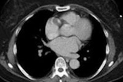
The use of an advanced iterative reconstruction technique with 3D processing can improve image quality and interreader agreement in reduced- and low-dose CT scans for patients with pulmonary diseases, Japanese researchers have found.
In a Web-exclusive article published in the October issue of the American Journal of Roentgenology, a team led by Dr. Yoshiharu Ohno of Kobe University Graduate School of Medicine found that the Adaptive Iterative Dose Reduction (AIDR) 3D technique produced significantly higher image quality scores on reduced- and low-dose CT scans compared to studies without the reconstruction method. In addition, interreader agreement in scans processed via AIDR 3D was determined to be nearly perfect.
"AIDR 3D was found to be useful for assessment of image noise reduction and radiologic findings on reduced- and low-dose CT for patients with various pulmonary diseases," the authors wrote.
Seeking to test their hypothesis that an AIDR 3D algorithm could yield improvements in image noise and reductions in radiation dose in low-dose chest CT examinations while maintaining diagnostic accuracy, the researchers studied 37 consecutive patients with pathologically diagnosed lung cancer and various pulmonary diseases (AJR, October 2012, Vol. 199:4, pp. W477-W485).
The patients included 17 men and 20 women with a mean age of 66 years (range, 35-84). There were 10 lung cancers, seven cases of asbestos-related pleural disease, five cases of organizing granulomas, four metastatic lung tumors, four thymomas, three granulomas, two cases of pulmonary tuberculosis, and one case each of cryptococcosis and giant bulla. In addition, 10 had pulmonary emphysema and eight had interstitial lung disease due to connective tissue diseases, according to the researchers.
All patients received chest MDCT exams on either an Aquilion 16 or 64 scanner (Toshiba Medical Systems) at three different tube current levels: 25, 50, and 150 mAs. The standard-dose (150 mAs) image datasets were reconstructed as thin-section CT without AIDR 3D (Toshiba), while both the reduced-dose (50 mAs) and low-dose (25 mAs) datasets were reconstructed as thin-section CT without and with AIDR 3D. All CT sets that were not reconstructed using AIDR 3D were reconstructed using filtered back projection (FBP).
Without access to the exams' technical parameters, a radiologist with more than 10 years of experience in chest CT and another board-certified chest radiologist with 21 years of experience then independently reviewed the MDCT images. The studies were randomized.
Image noise was quantitatively assessed with region-of-interest measurements, and readers were asked to evaluate the likelihood of occurrence of the following: emphysema, ground-glass opacity, reticular opacity, interlobular septal thickening, bronchiectasis, honeycomb pattern, nodules at the lung window setting, aneurysm, calcification at coronary artery, pericardial or pleural effusion, pleural thickening, pleural calcification, and lymphadenopathy at the mediastinal window setting. A five-point visual scale was used, with 1 meaning absent and 5 meaning present.
Differences in image quality
From quantitative assessments, the researchers determined that the image quality of both reduced- and low-dose CT without AIDR 3D was significantly lower than that of standard dose-CT and that of reduced- and low-dose CT with AIDR 3D (p < 0.05). Low-dose CT without the AIDR 3D method was also determined to be of significantly poorer image quality than reduced-dose CT without AIDR (p < 0.05).
In qualitative assessments, reduced- and low-dose CT without AIDR 3D were judged to have significantly lower image quality than standard-dose CT and also reduced- and low-dose CT with AIDR 3D (p < 0.05). Again, the researchers determined that low-dose CT without AIDR 3D produced image quality that was significantly poorer than reduced-dose CT without AIDR 3D (p < 0.05).
Interreader agreements for emphysema, honeycomb pattern, and nodules were almost perfect (κ > 0.81), while all agreements for ground-glass opacity and bronchiectasis were substantial or almost perfect (κ ≥ 0.81), according to the researchers. Furthermore, all agreements for reticular opacity and interlobular septal thickening were found to be moderate, substantial, or almost perfect (κ ≥ 0.72), respectively.
In assessing agreements between standard-dose CT and others on the mediastinal window setting, all agreements for mediastinal findings were almost perfect (κ > 0.81), the researchers found.
"According to our findings, we can reduce tube current from 150 to 25 mAs without significantly diminishing the image quality and capabilities of radiologic findings, except for detection of reticular opacity and interstitial septal thickening, in routine clinical practice," the authors wrote. "In addition, if we hope to assess all radiologic findings without any reduction in image quality or radiologic findings capability, we can reduce tube current to 50 mAs with the adaptation of AIDR for chest CT examination in routine clinical practice."
Acceptable for routine clinical use
Because AIDR 3D provides almost no reconstruction time penalty compared with protocols using the standard FBP algorithm, AIDR 3D can be considered acceptable for use in routine clinical practice, according to the researchers.
"We, therefore, think that AIDR is a useful algorithm for chest CT from the point of view of radiation dose and clinical practice issues," they wrote.
The authors noted that a prospective cohort study and a diagnostic performance study appear advisable to further evaluate the use of AIDR with low- and reduced-dose CT.
|
Study disclosures The study was supported by a grant from Toshiba and a Grant-in-Aid for Scientific Research from the Japanese Ministry of Education, Culture, Sports, Science, and Technology. |


