"Pleural effusions are a common finding on CT, and current practice is for the radiologist to estimate a size of the effusion once identified," noted co-author Dr. Matthew Moy, now a resident at Massachusetts General Hospital. "There are no published criteria to guide this estimation, and sizing can differ between radiologists."
What one considers small, another may think of as moderate, and this can lead to confusion for clinicians reading radiology reports, he said.
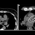
The study aimed to develop and validate a simple grading system that characterizes effusions as small, moderate, or large based on a variety of qualitative and simple quantitative CT features. These features were compared among 34 effusions of varying sizes measured using a volume segmentation tool.
The features that best correlated with effusion size were used to create the grading system used in the study. Moy and colleagues hoped that the grading system would improve interreader agreement among radiologists and clinicians.
The CT features that best divided effusions into small (< 20%), moderate (20% to 40%), and large (> 40%) effusions were anteroposterior (AP) quartile and maximum AP depth at the midclavicular line. The decision rule categorizes first AP quartile effusions as small, second AP quartile effusions as moderate, and third and fourth AP quartile effusions as large. The two-step decision rule improved interobserver agreement for pleural effusion quantification among various physicians, according to the study team.
"This grading system improves consistency among radiologists and clinicians, thereby improving communication of radiological findings," Moy said. "The grading system may also be useful in guiding management, such a


