Sunday, November 25 | 10:45 a.m.-10:55 a.m. | SSA19-01 | Room S403B
This scientific presentation will describe how computer-aided detection (CAD) software has the potential to increase the detection of acute vertebral body fractures on trauma CT studies.Radiology practices today face the challenge of dealing with growing workloads due to the increasing technical sophistication of CT scanners and declining reimbursement. At the same time, they are also dealing with increasing expectations of the general public and ordering physicians for rapid and error-free image interpretation, said co-author Dr. Joseph Burns, PhD, a former U.S. National Institutes of Health (NIH) Imaging Sciences Training Program fellow and currently an associate clinical professor at the University of California, Irvine.
The development of automated, secondary "shadow" readers that scan images in parallel with an interpreting radiologist could be a potential solution to the challenge of assessing large datasets in short time intervals, Burns said. Such a CAD system would be used by the radiologist as an assistant.
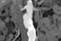
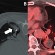
The NIH team sought to design and test novel methods for fully automated detection of thoracolumbar spine fractures on trauma CT studies. Its CAD system was able to detect and localize vertebral body fractures with low false-positive rates on spine CT studies.
Burns noted that computerized image analysis is not intended to replace radiologist interpretation of imaging studies.
"The intricate thought process complexity and situational plasticity of the human mind allows for recognition of arbitrary, innumerable a priori unexpected pathologies and adaptive image perception for varying geometric perspectives of the same objects," Burns said. "However, CAD can and will be a useful tool in the radiologists' armamentarium, acting as an automated assistant and error checker, designed to analyze images for specific, targeted, high-risk pathologies."


