A second-generation 320-detector-row CT scanner can significantly lower radiation dose during coronary CT angiography (CCTA) procedures while maintaining a high level of image quality, according to research published online in Radiology.
Researchers from the National Institutes of Health evaluated CCTA radiation exposure and image quality on a second-generation 320-slice CT system in more than 100 patients and found dramatic reductions in both median radiation dose and median size-specific dose estimates, as compared with a first-generation system.
"Minimizing radiation exposure while maintaining diagnostic-quality scans is clearly feasible with this new second-generation 320-detector-row CT scanner," wrote lead author Dr. Marcus Chen and colleagues. "The low dose achieved during CT angiography could be used to minimize overall radiation dose to the patient or to enable additional types of imaging (e.g., perfusion imaging) within reasonable radiation doses" (Radiology, January 22, 2013).
The researchers performed contrast-enhanced CCTA on an Aquilion One Vision CT system (Toshiba Medical Systems) for 107 adults with a mean age of 55.4. Imaging was performed with a gantry rotation time of 275 msec, and images were reconstructed with a 512 x 512 matrix, 0.5-mm-thick sections, and 0.25-mm increments using adaptive iterative dose reduction (AIDR) 3D software (Toshiba) and asymmetric conebeam reconstruction.
They then retrospectively compared the radiation exposure and image quality to those of CCTA exams previously performed on a separate cohort of 100 patients using a first-generation 320-slice scanner. The CT parameters were similar.
The researchers estimated effective radiation dose by multiplying the dose-length product by an effective dose conversion factor to produce size-specific dose estimates (SSDE). Two experienced cardiovascular imaging physicians evaluated image quality independently and in a blinded fashion.
|
In other findings, overall radiation dose was less than 0.5 mSv for 23 (21.5%) of the 107 CCTA exams, less than 1 mSv for 58 (54.2%), and less than 4 mSv for 103 (96.3%).
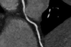
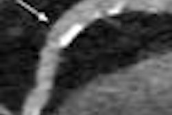
The readers reported that all studies were of diagnostic quality, with most having excellent image quality. The mean image quality score ranged from 3.63 ± 0.49 to 3.84 ± 0.42 on a scale of 1 (unevaluable, with severe artifacts rendering diagnostic interpretation impossible) to 4 (excellent, no significant artifact).
"The combination of a gantry rotation time of 275 msec, wide volume coverage, iterative reconstruction, automated exposure control, and larger x-ray power generator of the second-generation CT scanner provides excellent image quality over a wide range of body sizes and heart rates at low radiation doses," the authors concluded.

