In one of the first studies to evaluate advanced CT iterative reconstruction techniques in children, researchers found that model-based iterative reconstruction (MBIR) can halve pediatric CT radiation dose, delivering images of equal or better quality than standard-dose exams and outperforming first-generation IR software, according to a new article in Radiology.
In 25 young patients who had undergone standard-dose CT for a malignancy within the prior year, a second, reduced-dose CT scan cut radiation dose by an additional mean of 46%, as measured by CT dose index volume (CTDIvol). Even so, the MBIR images delivered significantly less noise and better spatial resolution than the standard-dose exams. Still, MBIR yielded inferior image quality in the bones, with better quality in the lungs and soft tissues. The 100% adaptive statistical iterative reconstruction (ASIR) images were inferior for all of the above.
"Both objectively and subjectively, image quality of reduced-dose images reconstructed with model-based iterative reconstruction is superior or equivalent to that of standard-dose CT images and is superior to that of reduced-dose CT images reconstructed with 100% adaptive statistical iterative reconstruction in pediatric patients undergoing body CT," wrote Dr. Ethan Smith, from the University of Michigan Health System's Mott Children's Hospital, and colleagues (Radiology, October 3, 2013).
MBIR vs. ASIR
MBIR (GE Healthcare) differs from other iterative reconstruction techniques in its ability to account for the optics of the CT scanner, including focal spot and detector size. And unlike ASIR's gradations, MBIR can only be applied in one strength. A few MBIR studies have been performed, but they have been mostly limited to adult subjects, the authors noted.
The study aimed to compare image quality and radiation dose between a reduced-dose CT protocol with MBIR and ASIR and a standard-dose CT protocol with 30% ASIR, they wrote.
"It's the first time this has ever been done in children; there have been a couple of studies of adults using MBIR and looking at dose reduction, but children are a little different," Smith told AuntMinnie.com. "Children tend to have less abdominal fat and less tissue that allows you to see organs and the contrast between things. The structures are smaller ... so it's usually harder to see things in children."
All 25 subjects were younger than 21 years of age (mean, 10.8 years) and undergoing follow-up CT for a known malignancy of the chest, abdomen, or pelvis on a 64-detector-row scanner (Discovery HD750, GE). Within the previous year, all had undergone standard-dose CT on the same scanner, but using 30% ASIR reconstruction.
Dose reduced 30%, then another 50%
The reduced-dose protocol for the MBIR exams used automatic tube current modulation with a fixed noise index of 47.7 -- a level specifically chosen for its ability to provide an approximate 50% dose reduction versus the standard exam noise index of 33.6, the study team wrote. Weight-based settings were used to determine exposure at 80, 100, or 120 kVp, but it was always set to the same level as the previous standard-dose exam. Oral and intravenous contrast in all exams was dosed using weight-based algorithms, according to the authors.
Scanner CTDIvol and phantom size were documented to ensure their appropriateness for size-specific dose estimate (SSDE) calculations assessed for each exam, the group wrote. The researchers calculated both objective image noise and subjective image quality based on review by two experienced radiologists.
"We went back and looked at ... how the image quality was; whether we were able to see all the lesions we wanted to see; subjectively, how did the images look; and we looked at a couple of objective measures: image noise and the standard deviation of the Hounsfield units of a given region of interest," Smith said. "Our physicist used the exact same scanning parameters and actually scanned a phantom and looked at the spatial resolution -- and the phantom [image] was superior to standard dose using the MBIR technique."
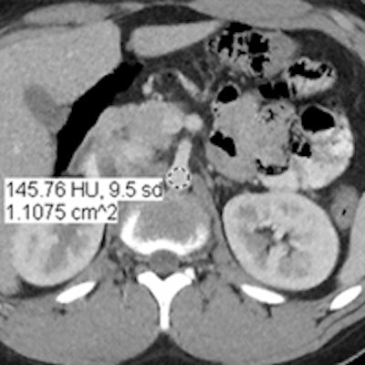
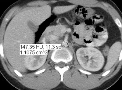
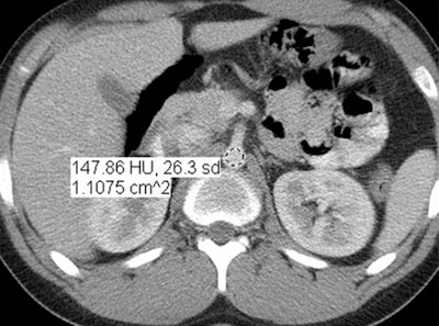
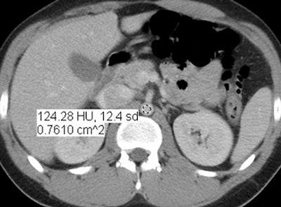
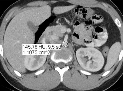 Images are of a 17-year-old boy with a history of Hodgkin's disease. Reduced-dose MBIR image (above), reduced-dose 100% ASIR image (below), reduced-dose filtered back projection image (third from top), and comparison standard-dose 30% ASIR image (bottom), all with a region of interest in the abdominal aorta for objective noise measurement. The reduced-dose MBIR image has significantly less noise compared with other reduced-dose and standard-dose reconstructions. Images republished with permission of RSNA from 10.1148/radiol.13130362, October 3, 2013.
Better at lower doses
In an objective evaluation, reduced-dose MBIR images had decreased image noise compared with standard-dose 30% ASIR images (for example, 12.7 HU versus 19.4 HU in the aorta, respectively, and 8.7 HU versus 14.2 HU in the liver).
Between the original images and the new images acquired using MBIR and the reduced-dose protocol, the mean decrease in CTDIvol was 46% (range, 19%-65%), with a similar decrease in SSDE of 44% (range, 19%-64%).
The mean CTDIvol for reduced-dose MBIR examinations was 3.1 mGy ± 2.3 (range, 1.0-11.3 mGy). Mean CTDIvol for comparison standard-dose examinations was 5.6 mGy ± 3.3 (range, 2.1-13.9 mGy). The wide range of patient sizes and ages made for significant variability, Smith said, but patient for patient, the MBIR doses were far lower.
"For example, our lowest dose in reduced-dose CT with MBIR was 1 mGy at SSDE compared to 2.2 mGy for the standard protocol," he said. The mean SSDE for reduced-dose examinations was 3.2 mGy ± 2.6 (range, 1.0-11.4 mGy), compared with 5.6 mGy ± 4.0 (range, 2.2-14.5 mGy) for standard-dose exams.
The MBIR images also had less noise (p > 0.004), and spatial resolution was superior overall for MBIR. The readers found MBIR images equivalent to standard-dose images for the lungs and soft tissues (p > 0.05) but inferior for bone imaging (p = 0.004), the group reported.
"The only place where the MBIR wasn't rated as equivalent to standard-dose images was when we looked at the bones," Smith said. "I think it was more the smoothing or 'plasticky' effect of it that made the trabeculation of the bone less apparent, so it almost looked more like smooth bone as opposed to being able to see the tiny trabeculae."
In current practice, the clinicians maintain the low MBIR dose but also routinely examine the native images, he said.
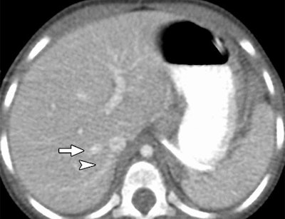
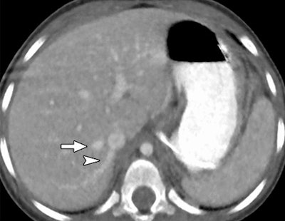 Reduced-dose axial CT images reconstructed with MBIR (above) and 100% ASIR (below) in a 30-month-old girl with a history of a malignant germ cell tumor in the right hepatic vein (arrow) and an accessory right hepatic vein (arrowhead). Images republished with permission of RSNA from 10.1148/radiol.13130362, October 3, 2013.
The sharper image
Image quality was also judged to be sharper with MBIR. Using the same reduced-dose acquisition, lesion detectability was better (32 of 84 rated lesions, 38%) or the same (52 of 84 rated lesions, 62%) with MBIR compared to 100% ASIR reconstruction.
CT imaging with the MBIR technique enables significant reduction of patient radiation dose while preserving diagnostic image quality, the authors concluded. By using the parameters described, both CTDIvol and SSDE measurements showed similar mean dose reductions of approximately 45%, compared with a previous low-dose acquisition that dropped the standard dose by one-third.
The study's major limitation was the lack of reviewer blinding, which was impossible due to the inherent differences in the appearance of MBIR versus ASIR schemes, the group wrote.
The strong findings have resulted in the implementation of low-dose MBIR imaging for all nonemergent outpatient studies at the hospital, Smith told AuntMinnie.com. Reconstruction times -- always an issue for MBIR -- have been reduced from 24 hours at an external location to about an hour per study onsite, but an hour is still too long for emergency radiology, he said. And for now, doses are being kept a little higher than the levels achieved in the study.
Despite its benefits, MBIR images do take some getting used to, he added.
"We were actually somewhat surprised to see the images look inherently different," he said. "ASIR was weird looking, and these also look weird, but when we look at them and ask, 'Are we seeing the lesions? Are we seeing the anatomic structures we need to see?,' they were actually as good as standard dose, even though we reduced the dose by 50%."


