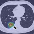
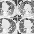
Quantitative assessment of tumors using dual-energy CT (DECT) can help predict the aggressiveness and invasiveness of lung adenocarcinoma, according to a study by researchers from South Korea that was presented at the RSNA 2013 meeting.
Analysis of DECT images using multiple factors -- from the presence of a solid component in a tumor to its uniformity on an iodine map -- could predict tumor grade before surgery takes place, the group found.
"With the recent imaging paradigm shift to personalized medicine, stratification of lesions is critical," said Jungmin Bae, from Samsung Medical Center, Sungkyunkwan University School of Medicine, in her RSNA presentation. "It is based on specific pathologic or genomic features and is expected to yield substantially good prognosis."
Traditional pathological indicators include tumor size, extent of invasion, lymph node involvement, and pleural and vessel invasion.
One problem is that 70% of lung adenocarcinomas are inoperable, which results in insufficient pathologic evaluation to determine when intervention might improve outcomes, Bae said.
Classification of lung adenocarcinoma pathologic subtypes has improved, though, with the 2011 publication of criteria from the International Association for the Study of Lung Cancer, the American Thoracic Society, and the European Respiratory Society (IASLC/ATS/ERS). These criteria calculate the extent of the invasive component; vascular, lymphatic, perineural, or pleural invasion; and the percentage of tumor cellularity.
Using these criteria, pathologic grade can be scored as follows:
- Score 1: Adenocarcinoma in situ, minimally invasive adenocarcinoma, lepidic
- Score 2: Acinar, papillary
- Score 3: Solid, micropapillary
Dual-energy CT can help determine which is which, according to Bae.
"Quantification using preoperative DECT imaging metrics can help to predict pathologic aggressiveness and invasiveness, which may help select the candidate for limited resection or adjuvant therapy," she said.
Usefulness of quantitative DECT
The prospective study aimed to evaluate the usefulness of quantitative analysis of DECT imaging metrics as predictors of histopathologic tumor grade and aggressiveness in lung adenocarcinoma.
The study team examined 60 patients (median age, 58) with biopsy-proven or radiologically suspected early-stage adenocarcinoma. Sixty-one percent of the patients were nonsmokers. All underwent DECT and PET/CT followed by surgery and pathologic tumor evaluation.
Pathologic evaluation was performed by two lung pathologists using virtual microscopy and virtual slides to apply IASLC/ATS/ERS lung adenocarcinoma classification criteria.
Out of a total 73 tumors, 47 (64%) were 2 mm or smaller, while 26 (36%) were larger than 2 mm. Nodal status was negative in 95% of patients.
Regarding the stage of adenocarcinoma, 48 (80%) of patients were stage IA, 10 (17%) were IB, and two (3%) were IIA.
Among the 73 tumors, four (5%) were adenocarcinoma in situ, 11 (15%) were minimally invasive adenocarcinoma, and 58 (80%) were invasive adenocarcinoma.
Multivariate analysis showed that the presence of a solid component, uniformity on an iodine map, tumor density, and reaching the 75th percentile CT attenuation value on noncontrast images were all statistically significant independent predictors of pathologic invasiveness.
As for tumor histologic grade, 33% of the tumors in the study were low grade, 53% were intermediate grade, and 7% were high grade.
| Association between tumor features and pathological grade | |||
| Variables | Low grade | Intermediate grade | High grade |
| Male-to-female ratio | 21:8 | 5:8 | 4:1 |
| Solidity: nonsolid/part-solid/solid | 23/2/4 | 10/14/5 | 0/1/4 |
| Maximum standardized uptake value (SUVmax) | 0.77 | 3.47 | 5.80 |
| Size -- lung setting (mm) | 17.53 | 25.26 | 24.0 |
| Size -- solid | 3.73 | 14.18 | 21.20 |
| Volume (cm3) | 3.30 | 8.09 | 8.65 |
| Mass (g) | 1.93 | 5.93 | 7.53 |
| Density | 0.45 | 0.74 | 0.89 |
"Lower grades tended to represent nonsolid and part-solid lesions," Bae said. "Among traditional factors obtained without sophisticated quantification, smoking habits, tumor solidity, and SUVmax were proven to be significant factors in defining tumor grade."
Several collections of variables were used to create a series of models in an effort to find the best predictors of pathologic tumor grade.
A multinomial logistic regression model using gender, SUVmax, skewness, and kurtosis showed the largest area under the curve (AUC) at 0.97 for discriminating low- from intermediate-grade solid tumors, and 0.95 for discriminating intermediate- from high-grade solid tumors.
The best-performing of these models, Model 7, showed highly significant improvement in discriminating tumor grade with the use of quantitative DECT parameters.
"Model 7 reveals that by adding to CT quantitative parameters, the area under the curve is significantly increased, with a p-value of less than 0.05 in both low grade and high grade," Bae said.
"Quantitative imaging using preoperative DECT imaging metrics can help predict histopathologic tumor grade and aggressiveness," Bae concluded.
