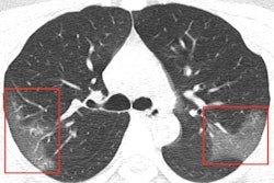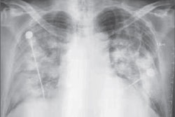
Researchers in Saudi Arabia have clarified defining characteristics of Middle East respiratory syndrome coronavirus (MERS-CoV) infection, which they say manifests in a similar way to organizing pneumonia on CT, according to a new report in the American Journal of Roentgenology.
Patients hospitalized with MERS most commonly showed bilateral predominantly subpleural and basilar airspace changes, with extensive ground-glass opacities and less consolidation, concluded the study team from King Abdulaziz University Hospital in Jeddah. The subpleural and peribronchovascular nature of most of the abnormalities is suggestive of an organizing pneumonia pattern, they wrote.
"A few studies have described variable degrees of lung opacities in patients with MERS, but did not clearly address their exact distribution," said lead author Dr. Amr Ajlan in a statement about the study. "Because we evaluated the CT findings in this laboratory-confirmed group of MERS patients, we had the ability to better characterize the nature and distribution of the abnormalities."
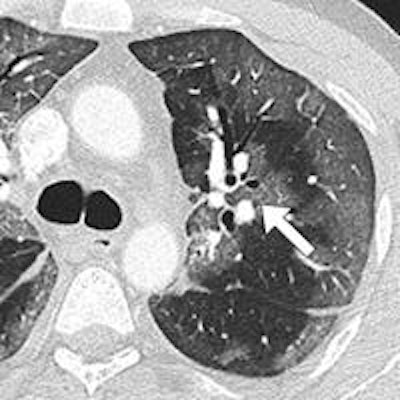
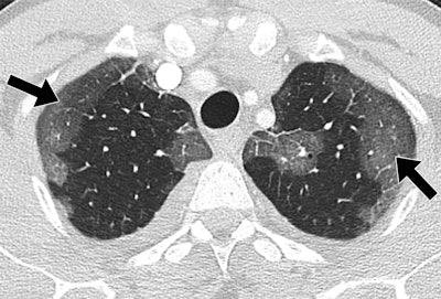 44-year-old man with MERS had no prior health problems. CT was performed one day after admission and nine days after symptom onset. The patient died in the intensive care unit. Upper-lung CT image shows large areas of bilateral subpleural ground-glass opacities. All images republished with permission of the American Roentgen Ray Society.
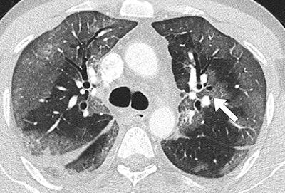 Midlung CT image shows that ground-glass opacities have peribronchovascular distribution (arrow).
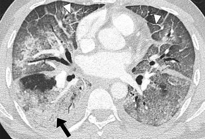 Lower-lung CT image shows more extensive and confluent basal abnormalities, with right lower lobe consolidation (arrow) and bilateral smooth interlobular septal thickening (arrowheads).
Several recent publications have examined MERS, but it has mostly been in nonimaging medical literature, and descriptions of the imaging features are sparse, especially in CT, the study authors wrote. What descriptions exist characterize the infection simply as bilateral patchy or extensive opacities (AJR, June 11, 2014).
The study aimed to provide detailed descriptions of chest CT findings in seven patients with MERS-CoV infection (five men and two women, ages 19-83 years). The patients were scanned with either a 16-slice CT scanner (Discovery CT750 HD, GE Healthcare), a 64-detector-row scanner (Somatom Sensation, Siemens Healthcare), or a 128-detector-row dual-source scanner (Somatom Definition Flash, Siemens). They were scanned at 100-120 kV and 200-500 mAs, using 1- to 2.25-mm slice thicknesses and 1.25-mm slice intervals. Only one patient received iodinated contrast (60 mL).
The images were interpreted by one of two fellowship-trained thoracic radiologists, who looked for ground-glass opacities, consolidation, cavitation, centrilobular nodules, tree-in-bud pattern, septal thickening, perilobular opacities, reticulation, architectural distortion, subpleural bands, traction bronchiectasis, bronchial wall thickening, intrathoracic lymph-node enlargement, and pleural effusions.
The images showed that airspace opacities were more common than interstitial changes, according to the authors. Five patients had both ground-glass opacities and consolidation -- with ground-glass opacities being more extensive in all but one of those patients. One patient had only ground-glass opacities and another had isolated consolidation.
The radiologists found smooth septal thickening in three of the seven patients, and minimal peripheral reticulation, traction bronchiectasis, and perilobular opacities were found in just one patient. None of the patients had tree-in-bud pattern, cavitation, or intrathoracic lymph-node enlargement, the authors wrote.
The most common CT finding in hospitalized patients with MERS is bilateral predominantly subpleural and basilar airspace changes, with more extensive ground-glass opacities than consolidation, Ajlan and colleagues concluded.
"The predilection of the abnormalities to the subpleural and peribronchovascular regions is suggestive of an organizing pneumonia pattern," they wrote. "Recognizing this pattern in acutely ill patients living in or traveling from endemic areas may help in the early diagnosis of MERS-CoV infection."


