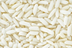
DUBAI - A veritable gastronomic feast was served up at this week's Total Radiology Conference at the multidisciplinary Arab Health show, when a presentation worthy of a Michelin star was delivered about food-inspired signs in radiology.
"Recognition of these signs, served with an understanding of the relevant differential diagnosis and pathologies, will go a long way toward satisfying the radiologist's ever-present hunger for learning and thirst for knowledge," said Fathima Hasan Mohamed, CT radiographer at the Clinical Imaging Institute, Al Ain Hospital in Al Ain, United Arab Emirates.
The radiologic signs of many diverse conditions and diseases have classic manifestations that resemble various types of food, she explained. These descriptive food signs are highly memorable and easily recognizable, and allow a confident diagnosis on the basis of imaging findings alone or can help narrow down the differential diagnosis.
Mohamed thinks the following signs are well worth looking out for and getting to know:
-
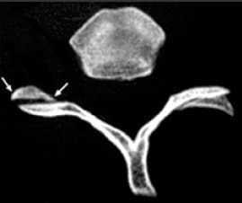
 On CT, the vertebral facet joint space can look like a hamburger. All images courtesy of Fathima Hasan Mohamed.
Hamburger: "My own personal favorite," Mohamed commented. On axial CT, the vertebral facet (apophyseal) joint space can look like a hamburger. When the facet joint is dislocated, the articular facets become uncovered, or naked, and this is also called naked facet sign. This CT sign is characteristic of a flexion-distraction injury and indicates severe ligamentous disruption and spinal instability. It may be unilateral or bilateral, depending on the facet dislocation.
Sausage digit: Refers to the appearance of diffuse fusiform swelling of digits due to soft-tissue inflammation from underlying arthritis or dactylitis. The most common cause is psoriatic arthropathy, but other causes include osteomyelitis, sickle cell anemia, sarcoidosis, and tuberculous dactylitis (spina ventosa).
Apple core lesion: The annular carcinoma of the colon produces focal circumferential thickening of the bowel wall and narrowing of the colonic lumen, associated with shouldering. On a barium exam, the affected colon looks like a partially eaten apple.
Berry aneurysm: On CT, the intracranial saccular aneurysm may resemble the size and shape of a berry. They are congenital and characteristically occur where arteries bifurcate; they may occur in isolation or in association with polycystic kidneys or coarctation of the aorta.
Banana: The banana appearance of the cerebellum on antenatal ultrasound is due to downward traction of the spinal cord and brainstem in neural tube defects. It is also seen in most fetuses with spina bifida but disappears after 24 weeks. Often there is concurrent hydrocephalus.
Lemon: The appearance of the fetal cranial vault on antenatal ultrasound resembles a lemon, and this is an excellent indicator of open spina bifida. An abnormally shaped fetal head on a second-trimester scan performed can resemble a lemon in the axial cross section because the frontal bones are flattened or concave.
Celery stalk metaphysis: Refers to vertical striations in the metaphysis of long bones or pelvis produced by a dysplastic bone disease such as osteopathia striata. Similar presentation may be seen in congenital rubella infection.
Onion skin periosteal reaction: In an aggressive bone lesion such as acute osteomyelitis, Ewing's sarcoma, or osteosarcoma, the successive deposition of periosteal layers gives a lamellated or onion skin appearance on plain films.
Coffee bean: This characteristic sign of sigmoid volvulus consists of a greatly distended, air-filled loop of sigmoid colon extending from the pelvis on abdominal radiography. The medial walls of the dilated bowel form a distinct oblique line that resembles the cleft of a coffee bean. It arises from the pelvis and may be large, with its apex often extending above the level of T10 to the left or right of the midline.
Eggshell calcification: Consists of shell-like calcification in the periphery of a node and occurs in up to 5% of cases of silicosis. Hilar nodes are predominantly involved, but mediastinal, cervical, and intraperitoneal nodes also may be affected. This calcification also has been observed following treatment for lymphoma, appearing one to nine years after radiation therapy. It may occur in patients with sarcoidosis and is usually associated with advanced pulmonary disease, as well as coal workers' pneumoconiosis, amyloidosis, and scleroderma.
Honeycomb lung: Refers to an advanced stage of fibrosis in which normal lung parenchyma is replaced by cystic spaces of 5 mm to 10 mm. It is visible on a chest x-ray and CT. The common causes are pneumoconiosis, sarcoidosis, fibrosing alveolitis, scleroderma, and rheumatoid diseases.
Linguine: After implantation of a silicone or saline breast implant, a fibrous capsule or scar forms around the implant shell. In an intracapsular rupture, the contents of the implant are contained by the fibrous scar, while the shell appears in a group of wavy lines. The linguine sign is most commonly picked up on MRI, and the same findings are seen on CT in patients with bilateral ruptured saline implants.
Doughnut: Refers to circumferential thickening of the bowel wall in carcinoma of the colon and inflammatory bowel disease.
Licked candy stick appearance: Refers to tapering of the tips of the metacarpal and metatarsal bones, phalanges, or clavicles, and is associated with psoriatic arthropathy, rheumatoid arthritis, and leprosy.
Cottage loaf: MRI appearance of the partially herniated liver through the ruptured right hemidiaphragm.
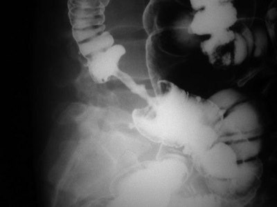 Double-contrast barium enema shows apple core lesion due to carcinoma of colon.
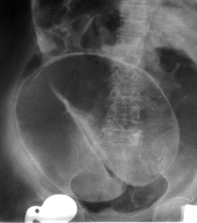 Coffee bean sign with perforation.
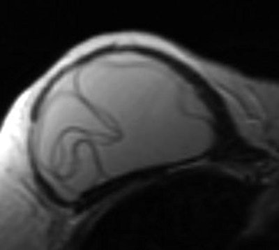 Breast MRI showing the linguine sign indicating intracapsular implant rupture.
"It may sound a little silly, but the radiology literature is replete with signs, some more fanciful than others," she concluded, before receiving an enthusiastic round of applause.

