Rice bodies in musculoskeletal (MSK) imaging are best seen on fluid-sensitive MRI sequences, and they can get bigger, or perhaps coalesce, over time and may disappear without interventional treatment, according to award-winning research from Turkey.
Rice bodies consist of small fibrinous bodies that are likely to represent the sequelae of microvascular infarcts of the synovium in the setting of chronic inflammation, but their pathogenesis and significance in articular tissue response to inflammation are still unclear, explained lead author Dr. Üstün Aydıngöz, a professor of radiology at Hacettepe University School of Medicine in Ankara.
These bodies are a unique feature of MSK radiology, and they have macroscopic similarity to polished grains of rice, he noted in an e-poster presentation at RSNA 2016 that received a certificate of merit when the poster hall judges revealed their winners on Wednesday afternoon.
In general, rice bodies originate from the synovium, and are almost exclusively seen where synovium is present, particularly in the joints, tendon sheaths, and bursae.
"The two conditions where there is abundance of small bodies within a synovium-lined cavity constitute the major differential diagnostic considerations: synovial (osteo)chondromatosis and lipoma arborescens," Aydıngöz stated. "However, synovial chondromatosis may be indistinguishable from rice bodies on MRI."
Rice bodies may be encountered in patients with chronic synovitis, either primary or reactive in nature, such as rheumatological diseases, osteoarthritis, tuberculous arthritis, and neuropathic arthritis. They can be seen with rheumatoid arthritis, juvenile idiopathic arthritis, seronegative inflammatory arthritis, osteoarthritis, tuberculous arthritis/tenosynovitis: tuberculosis, neuropathic arthropathy. Their nonvisualization on T1-weighted images helps differentiate them from other conditions.
"Rice bodies may undergo morphologic change over time. They may get bigger -- perhaps by coalescing," he pointed out. "Rice bodies may disappear without interventional treatment."
In these cases, it's also important to bear in mind the so-called horseshoe abscess. Paronas space is a potential space located at the level of the distal forearm lying between the flexor digitorum profundus tendons and the pronator quadratus fascia, and it is at this location where the radial and ulnar bursae can communicate (such communication was found in anatomical studies in 33% to 100% of patients). Tendon sheath infections originating in the thumb or little finger can follow this communicating pathway, resulting in the rare horseshoe abscess, according to Aydıngöz.
Turkey presents session
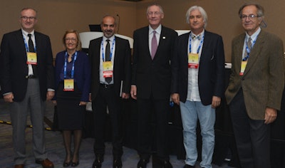
Leading radiologists from Turkey also featured on Tuesday in a special RSNA session, "Turkey Presents: The Meaning of Evolution for Radiology and Advances in Neuroradiology" (see photo).
 Speakers and moderators during Tuesday's "Turkey Presents" session. From the left are Drs. Muzaffer Basak, Ayse Nur Oktay Alfatli, Tamer Kaya, RSNA 2016 President Richard Baron, Civan Islak, and Kamil Ugurbil.
The co-authors of the rice bodies exhibit were Drs. Zeynep Maraş; Özdemir, İnönü University School of Medicine, Malatya; Dr. H. Nursun Özcan, Dr Sami Ulus Children's Hospital, Ankara; and Altan Güneş and Fatma Bilge Ergen, both from Hacettepe University School of Medicine, Ankara.
For further reading on the topic, the authors recommend the following:
Forse CL, Mucha BL, Santos ML, Ongcapin EH. Rice body formation without rheumatic disease or tuberculosis infection: a case report and literature review. Clin Rheumatol. 2012; 31:1753-1756.
Edison MN, Caram A, Flores M, Scherer K. Rice body formation within a peri-articular shoulder mass. Cureus. 2016; 8(8):e718.
Huang CC, Ko SF, Weng LH, et al. Sonographic demonstration of hyperechoic fibrin coating of rice bodies in trochanteric bursitis: the "fried rice" pattern. J Ultrasound Med. 2006; 25:667-670.
özcan HN, Aydıngöz ü, Marangoz S, Kara A, Ergen FB. Rice bodies within the neuropathic hip in a child with congenital insensitivity to pain. Pediatr Radiol. 2015; 45:782-783.
Tan CH, Rai SB, Chandy J. MRI appearances of multiple rice body formation in chronic subacromial and subdeltoid bursitis, in association with synovial chondromatosis. Clin Radiol. 2004; 59:753-757.
Fairbairn N. A rare case of communicating infection in the hand: the horseshoe abscess. Injury Extra. 2012;43:25-27.


