Patients who received contrast with their CT scans had more than twice as many double-strand breaks in their DNA from radiation exposure compared to those who had CT studies without contrast, concludes a new study in the June edition of Radiology. But whether this DNA damage translates into cancer risk is unclear.
The study of 245 patients scheduled for chest CT exams (one-third acquired with contrast) examined blood samples before and after scanning, measuring damage to peripheral blood lymphocytes using fluorescence microscopy. After the scans, patients who had received contrast media showed more than 100% greater DNA damage than patients whose scans were unenhanced (Radiology, June 2015, Vol. 275:3, pp. 692-697).
"Our study showed that the application of iodinated contrast agents during chest CT scanning clearly increases the amount of peripheral lymphocyte DNA radiation damage," lead author Dr. Eike Piechowiak and colleagues wrote. "We know that DNA radiation damage causes cancer; therefore, if this damage is enhanced, the likelihood of cancer generation is theoretically increased."
Contrast media and DNA
Contrast is used in slightly more than half of all CT exams. Although the nephrotoxic and cytotoxic effects of contrast are well-known, DNA damage has not been part of the risk calculation in daily practice. However, one small study has already shown that contrast agents can increase radiation-induced DNA damage after CT or angiography, the group noted.
The current study aimed to evaluate the effects of iodinated contrast on the development of DNA double-strand breaks in patients undergoing diagnostic chest CT scans in a larger population than has been previously studied, wrote Piechowiak and colleagues Dr. Jan-Friedrich Peter; Beate Kleb; Dr. Klaus Klose, PhD; and Dr. Johannes Heverhagen, PhD, from Philipp University of Marburg in Germany and the University Hospital of Bern in Switzerland.
The study included 245 patients with a mean age of 64 years: 179 patients were scheduled for contrast-enhanced CT exams of the chest and 66 were scheduled for noncontrast chest CT scans.
All patients were examined on a dual-source CT scanner (Somatom Sensation 64, Siemens Healthcare). The contrast-enhanced group received an average 62 mL of contrast (Ultravist, Bayer HealthCare Pharmaceuticals) before the scans, which were acquired at 120 kVp with 0.5-sec rotation time and a 512 x 512 matrix. As for dose measurement, dose-length product (DLP) was recorded for each patient.
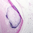
To measure DNA damage, the researchers used fluorescence microscopy to look for foci of a phosphorylated histone known as yH2AX. Measuring these foci has been well-established as a surrogate for calculating the number of DNA double-strand breaks induced by radiation damage.
To correct for any differences in radiation dose between individual scans, the number of yH2AX foci was standardized to a reference dose of 312 mGy-cm to reflect the reported linear relationship between foci per cell and dose.
The researchers acquired blood samples immediately before and after all CT exams. Blood lymphocytes were separated out, washed, and processed. The investigators used a fluorescence microscope equipped with a charge-coupled device camera and AxioVision software. For the quantitative analysis, the yH2AX foci were counted by eye with original magnification (630x).
The investigators then looked for differences between the number of foci that developed in the presence and absence of the contrast agent.
Increase in DNA damage
The two patients groups showed no significant differences (p > 0.05) in patient characteristics; however, mean radiation doses were higher on a statistically significant basis for the noncontrast-enhanced scans (342 mGy-cm) versus the contrast-enhanced scans (301 mGy-cm).
Before the scans, the two groups showed similar levels of yH2AX foci per cell, with 0.073 foci per cell for the noncontrast patients and 0.071 for the contrast-enhanced group (p = 0.94).
Levels of γH2AX foci were higher in both groups after scanning, but the patients who underwent contrast-enhanced CT showed an additional 0.056 foci per cell; this was 107% higher than the change in patients who underwent unenhanced CT, who had a mean increase of only 0.027 foci per cell.
"The application of iodinated contrast agents during diagnostic x-ray procedures, such as chest CT, leads to a clear increase in the level of radiation-induced DNA damage as assessed with γH2AX foci formation," the authors concluded.
The increased damage with contrast enhancement is likely caused by the generation of additional secondary electrons when the contrast material absorbs x-rays, they noted.
"Because of their high density, iodinated contrast agents absorb more x-rays than do human soft tissues," the group wrote. "In addition, the generation of secondary electrons is strongly dependent on the density of the absorbing material. Thus, these effects are synergistic, and the generation of secondary electrons is even more pronounced. These secondary electrons could potentially be the major cause of x-ray-induced DNA damage."
Potential study limitations include bias resulting from the disease state of the patients, and the fact that DNA damage in blood cell lymphocytes may not occur the same way in solid organs, according to the authors. And, of course, the relationship between DNA breaks and cancer risk is unknown.
Editorial concurs
In an accompanying editorial (pp. 627-629), researchers from the U.S. National Cancer Institute noted that the study was the largest of its kind to date. But they cautioned about drawing too many conclusions from it; while γH2Ax can be used to measure double-strand breaks in DNA, Piechowiak et al refrained from linking the findings to cancer risk, noted Amy Berrington de Gonzalez, PhD, and Ruth Kleinerman.
In any case, one must also consider the other risks associated with contrast use, including allergic reactions and nephrotoxicity, they wrote. They recommended using American College of Radiology (ACR) Appropriateness Criteria to determine in which clinical scenarios contrast should be used.
"Given the expanding evidence about the effect on dose and potential cancer risk, further research into patterns of use and the potential adverse effects of contrast material use with CT now seems warranted," Gonzalez and Kleinerman concluded.

