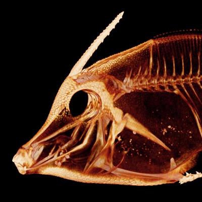
Is it possible to scan all the fish species in the world, store the data, and give it all away for free? One University of Washington professor believes it is -- and he's come up with some novel scanning techniques to move the project forward.
Adam Summers, PhD, a professor of biology and aquatic and fishery sciences, is behind the ambitious project to scan all species of fish and catalog them in an open-access database. Other researchers and institutions also are contributing their own data, and anyone is welcome to use the information collected. Looking forward, the hope is to eventually include all vertebrates.
"The idea of the database is brand new; it's completely unusual," Summers told AuntMinnie.com. While other places are offering smaller amounts of CT data, "what we're trying to do is put up everything that anyone might need for free, for whatever they want to do, whether it's science, education, or commercial use."
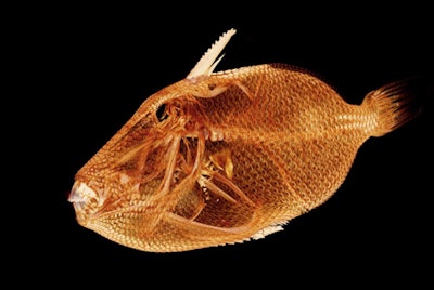
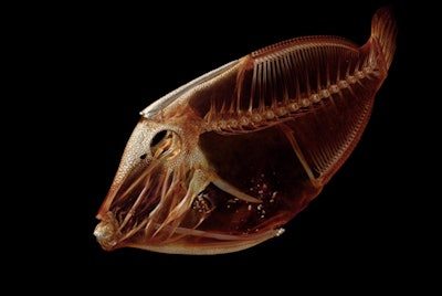 Above, Cantherhines sandwichiensis. Below, Trichomycterus maculatus. All images courtesy of Adam Summers, PhD.
Summers is head of the Comparative Vertebrate Biomechanics Lab at the university's Friday Harbor Laboratories on San Juan Island, about 80 miles from Seattle. He has been scanning fish for nearly 20 years, but initially it was catch as catch can, relying on CT scanners at other facilities. After obtaining a micro-CT scanner for the lab, he began scanning specimens and posting the images on Twitter.
"I would get this response from people, saying, 'Oh my gosh, what's coming next?' " he said. "And I would just say, 'I'm scanning them all' " -- with the goal of scanning as many fish as possible.
Workflow refinements
Workflow efficiency is a major consideration when scanning human patients, and, ironically, scanning fish is no different. After a couple of months of working with the micro-CT scanner (SkyScan 1173, Bruker), Summers realized that he could streamline the process, coming up with a method to scan approximately 10 to 15 fish per session, rolling them up together like a burrito. This greatly reduces the number of scans needed, making it much more feasible to scan the tens of thousands of fish species.
"For anyone who runs a CT scanner, they know that when you stick a person, or a pet, or a museum specimen in a scanner, everything inside the scan field is scanned, not just the skeleton that you're interested in," he said. "There's no technological reason why you want to scan a whole bunch of air, and from my point of view, every time we scanned air that was a huge waste."
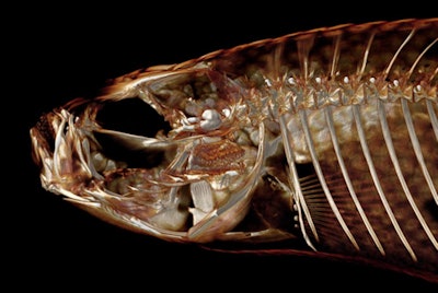 Above, Orestias sp. Below, Pervagor melanocephalus.
After a burrito of fish is scanned, Summers and colleagues use segmentation software from Bruker to isolate each fish. Next, they examine the data using either 3D Slicer or Horos open-source image visualization software.
The results can lead to surprising insights. For example, Summers described how the image of a piranha led to an understanding of their behavior. The particular fish had unusual damage to its skeleton, the shape of which led him to realize that it must have occurred from another fish biting it. These fish live in highly social environments, he said, and he suddenly realized that their main method of "chatting" with each other must be to nip -- "and when you have those kinds of teeth, nipping at each other is a big deal."
"If you look at their skeleton, I suddenly just saw it in a new light, and saw how much reinforcing there is," he said. "It's a really neat way to suddenly look at these fish."
Summers has also been printing 3D models from scans for many years, which can help with visualization of different species and their anatomy. Recently, for example, he printed the head of a 2-cm fish so that it was larger than his hand.
"We've always used [3D printing] to make little devices, and to make little anatomy big and big anatomy little, and now we just have a lot more data that we can print out," he said.
 Above, Rhinecanthus aculeatus. Below, Trinectes maculatus.
Hundreds of fish species have been scanned so far, with a Google Doc on the project's Open Science Framework site listing what's been accomplished to date, along with CT parameters and other information. Others are welcome to come to the island to scan their specimens for free, or they can send their items to the lab to be scanned, according to a release from the university. The fish must come from museum collections, however.
And while there are plenty of fish in the sea, so to speak, there are more subjects on the horizon. The project has led to a new initiative -- called oVert, or open vertebrates -- in which David Blackburn, PhD, from the University of Florida is organizing resources to obtain scans of all vertebrates, Summers said.

