Tuesday, November 29 | 11:00 a.m.-11:10 a.m. | SSG12-04 | Room S403B
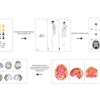
In this presentation, researchers will discuss the added value of spectral CT and k-edge imaging in the assessment of stented vessels to reduce artifacts.In conventional CT, the diagnosis of stented coronary arteries is limited due to strong blooming artifacts that often impair visualization of the vessel lumen. In comparison, spectral photon-counting CT (SPCCT) with a multibin prototype scanner benefits from increased spatial and spectral resolution.
The study used both in vivo (animal model) and in vitro imaging to evaluate stents made of platinum, cobalt-chromium, or stainless steel. Gadolinium was used as the contrast agent. All image reconstructions used an in-plane pixel size of 0.2 x 0.2 mm2 for image reconstructions.
"The increased spatial resolution of SPCCT resulted in a significant reduction in blooming artifacts that improved vessel lumen delineation and improved stent metallic mesh visualization," Monica Sigovan, PhD, from the University of Lyon in France, told AuntMinnie.com. The technique offered other advantages for isolating and visualizing the stent lumen as well.
"This high-resolution stent-specific imaging may allow validation of correct stent deployment and detection of mesh integrity problems," Sigovan said.


