Sunday, November 26 | 10:55 a.m.-11:05 a.m. | SSA05-02 | Room S404CD
The use of 4D CT scans may boost radiologists' accuracy for detecting pleural invasion in lung cancer, according to researchers from Japan.High radiation dose is a concern associated with 4D CT that has somewhat deterred its regular use by radiologists. However, the notable improvements that 4D CT offers in terms of diagnostic accuracy may warrant its use in certain scenarios, Dr. Tsuneo Yamashiro from the University of the Ryukyus in Nishihara told AuntMinnie.com.
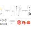
The researchers assessed the diagnostic accuracy of two chest radiologists in determining the presence or absence of pleural invasion, which the researchers subsequently confirmed via surgery. The 60% sensitivity and 77% specificity using conventional chest CT scans fell short of the 100% sensitivity and specificity provided by the 4D CT scans when combined with a colored map.
In a prior study, the researchers demonstrated the potential for 4D CT to serve as an alternative approach for the preoperative diagnosis of parietal pleural invasion of lung cancer, according to Yamashiro (European Journal of Radiology, February 2017, Vol. 87, pp. 36-44). Now, they found that using software to automatically assess the enhanced visualization afforded by 4D CT markedly improved diagnostic accuracy, compared with conventional CT.
"Our ultimate goal is not just making a correct diagnosis for parietal pleural invasion and adhesion by lung cancer," he said. "Our true goal is to detect benign (inflammatory) adhesions between the parietal and visceral pleura, which often cannot be found on conventional chest CT."


