Tuesday, November 28 | 12:45 p.m.-1:15 p.m. | VI175-ED-TUB11 | Lakeside, VI Community, Station 11
In this poster presentation, researchers will discuss how 3D printing facilitates planning for endovascular intervention, especially for replacing aortic valves.Prior research has repeatedly substantiated the usefulness of 3D printing for planning various operations, including hip surgery and stroke clot removal. In recent years, physicians have elected to perform transcatheter aortic valve implantation more often than traditional surgical replacement to manage faulty aortic valves.
"By combining the two new modalities of 3D printing and transcatheter aortic valve implantation, we have found that candidates for aortic valve replacement can derive tremendous benefits," Dr. Fernando Gutierrez from Washington University School of Medicine told AuntMinnie.com.
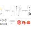
Gutierrez and colleagues used the CT scans of 20 patients deemed candidates for transcatheter aortic valve implantation to produce life-size 3D-printed models of the patients' aortic root. The researchers will present how their measurements from the 3D models compared with those made on data drawn from CT scans.
Prepping for the surgery with 3D-printed models enables cardiologists to choose the best type and size of implants, as well as determine the optimal positioning of the C-arm to minimize radiation exposure and reduce procedure time, according to the researchers.
"Our group has been continuing to develop pertinent protocols so that preplanning utilizing 3D printing can be performed in an expedited fashion," Gutierrez said.
The researchers intend to continue their investigation by expanding the patient base.


