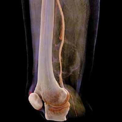
Cinematic rendering proved to be as accurate as conventional volume rendering and CT angiography (CTA) for presurgical identification of the potential invasion of soft-tissue tumors into major blood vessels, according to an article published online March 27 in the Journal of Computer Assisted Tomography.
The group, led by senior author Dr. ZhenHui Li, PhD, from Kunming Medical University in China, explored the possible utility of cinematic rendering in the evaluation of soft-tissue sarcomas, which are rare tumors that are challenging to diagnose due to their high malignancy rate and heterogeneous composition.
Sarcomas residing deep within soft tissue of the extremities have a tendency to invade major blood vessels and, thus, eliminate treatment options such as limb-saving surgery, the authors noted. Clinicians commonly rely on MRI and CT scans to evaluate tumor invasion before deciding on the best course of treatment, but these imaging modalities offer a rather limited view of the spatial relationship between tumors and adjacent vessels.
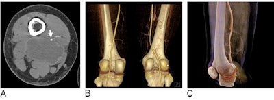
Seeking alternative visualization methods to confirm tumor invasion, Li and colleagues acquired the CTA scans of 43 patients who presented to the hospital with a deep soft-tissue sarcoma in one of their extremities between January 2013 and June 2016. The researchers reconstructed these scans with two techniques: traditional 3D volume rendering and cinematic rendering (syngo.via VB10, Siemens Healthineers).
 Visualization of a soft-tissue myofibroblastoma near the femoral artery of a 25-year-old woman using CT angiography (A), traditional volume rendering (B), and cinematic rendering (C). Image courtesy of Li et al. CC BY-NC 4.0.
Two musculoskeletal radiologists with at least nine years of experience examined the imaging data from CTA and the two advanced visualization techniques. Approximately 58% of the patients were male, and their average age was 49. In all, 21% of patients had tumor invasion based on the reference standard, the confirmed presence or absence of tumor invasion during surgery.
The radiologists were able to identify tumors invading major vessels on the cinematically rendered CT scans with nearly the same accuracy as on the CTA and volume-rendered scans. There were no statistically significant differences between the three techniques.
| CTA, volume rendering, and cinematic rendering for detecting sarcoma invasion | |||
| CT angiography | Volume rendering | Cinematic rendering | |
| Area under the curve | 0.92 | 0.86 | 0.77 |
| Accuracy | 0.77 | 0.77 | 0.70 |
| Sensitivity | 0.89 | 0.89 | 0.78 |
| Specificity | 0.75 | 0.75 | 0.68 |
| Positive predictive value | 0.47 | 0.47 | 0.39 |
| Negative predictive value | 0.96 | 0.96 | 0.92 |
In addition to matching the accuracy of the other methods, cinematic rendering provided a longer longitudinal view of the vessels and more photorealistic anatomic details, which helped optimize tumor visualization, according to the authors.
Though not statistically significant, the slightly lower sensitivity and positive predictive value of cinematic rendering may point to possible disadvantages of the technique, Li and colleagues noted. On the other hand, these variations likely stem more from the radiologists' minimal experience in examining cinematically rendered CT scans than a deficiency in the technique per se.
"Therefore, radiologists should receive training for identification of the relationship between tumors and adjacent major vessels on [cinematic rendering] before the technique can be used in clinical practice," they wrote.


