Cinematic rendering allowed radiologists from Johns Hopkins University to uncover key features of spleen anatomy and pathology not evident on conventional CT scans -- carving out yet another niche role for the technique, which they detailed in an article recently published online in Diagnostic and Interventional Imaging.
At their institution, Dr. Elliot Fishman; Dr. Steven Rowe, PhD; and colleagues have explored the possible advantages of incorporating cinematic rendering -- a 3D visualization technique characterized by highly photorealistic detail and shadowing -- into the evaluation of aortic complications and gastric and renal pathologies, among numerous other clinical applications.
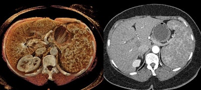 B-cell lymphoma of the spleen on a CT scan (right) and a cinematically rendered CT scan (left). All images courtesy of Dr. Elliot Fishman.
"Although the potential role of [cinematic rendering] in diagnostic imaging will still require a great deal of study to fully understand, the technique has shown promise in evaluating complex anatomy and pathology involving a number of regions of the body," they wrote (Diagn Interv Imaging, March 28, 2019).
 Cinematic rendering of an abdominal CT scan.
In the current article, the group discussed three distinct ways in which cinematic rendering could facilitate the evaluation of a variety of conditions affecting the spleen, including the following:
- Vascular conditions: Cinematic rendering is particularly well suited for visualizing vasculature near the hilum of the spleen, an area frequently affected by stomach and pancreatic tumors that restrict blood flow to certain parts of the spleen. Cinematically rendered CT scans display clear demarcations between these infarcted regions and otherwise stable areas.
In addition, the advanced visualization technique clearly depicts textural changes and fluid leakage in the spleen commonly associated with lacerations in trauma patients. These findings may additionally be able to help predict a patient's need for splenectomy.
- Neoplastic processes: The intrinsic ability of cinematic rendering to accentuate texture enables it to reveal spleen lymphomas, the organ's most common malignancy, more readily than standard CT -- thereby facilitating cancer diagnosis and prognosis, the authors noted. For rare tumors such as littoral cell angiomas, cinematic rendering can pinpoint underlying abnormalities that may help physicians distinguish between benign and malignant cases.
- Accessory spleen: Accessory splenic tissue seen on CT scans is occasionally mistaken for a tumor, especially for cancer patients. Standard CT and volume-rendered CT scans are often inadequate for determining tissue type in these cases and require a follow-up nuclear medicine exam to make a definitive diagnosis.
But cinematic rendering may obviate the need for additional testing in such cases because of the high level of soft-tissue detail it provides, as well as the pronounced differences it shows between the texture of tumor and normal tissue, according to the authors. "There may be tremendous opportunity to utilize this methodology to improve lesion characterization by visualization of texture within tumors and accessory splenic tissue," they wrote.
Cinematic rendering may provide surgeons with images that "better reflect intrinsic spatial relationships of structures within regions of complex anatomy," improve anatomical understanding for medical students and residents, bolster patient engagement, and even refine deep-learning algorithms, Fishman and colleagues wrote.
"However, at this time these potential applications of [cinematic rendering] remain speculative and much of the research that will underpin the use of [cinematic rendering] must still be undertaken," they concluded.


