A new biomechanics technique can estimate the long-term prognosis of abdominal aortic aneurysms (AAA) after surgical repair by analyzing CT angiography (CTA) scans, according to an article recently published online in Frontiers in Bioengineering and Biotechnology.
The current process for quantitatively assessing AAAs is to measure their maximum diameter on 2D CTA scans, where diameters greater than 5 mm are considered at risk of rupture. However, this diameter-based approach is unable to account for the significant variability that is common in the progression of AAAs and is, thus, not suitable for many cases, noted the authors, led by Karen López-Linares, PhD, from Pompeu Fabra University in Barcelona, Spain.
To address this limitation, the researchers sought to develop a more comprehensive approach to AAA rupture risk assessment using biomechanical strain analysis. They tested their method on a cohort of 22 patients who underwent endovascular aneurysm repair for an AAA followed by CTA scans at one and 12 months after surgery (Front Bioeng Biotechnol, November 1, 2019).
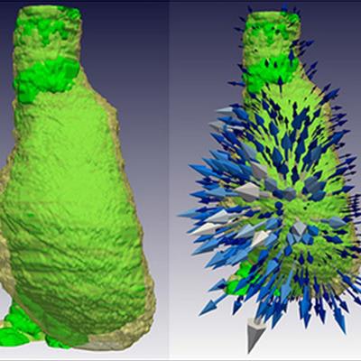
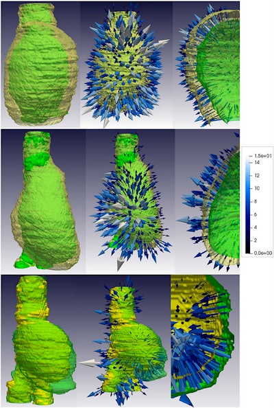
The group registered the CTA scans acquired at different time points to each other and then used biomechanical strain analysis to calculate the tensile and compressive strain on the aneurysms. The researchers subsequently generated 3D models of the AAAs displaying information from the strain analysis.
 Biomechanical strain analysis of patient CTA scans. Aneurysms with an unfavorable prognosis are depicted as 3D models with arrows pointing away from the aorta (top, middle). The arrows point toward the aneurysm when indicating a favorable prognosis (bottom). Image courtesy of Pompeu Fabra University.
On average, the technique yielded an area under the curve of 88.6% for estimating the patients' long-term prognosis.
"This suggests that the descriptors directly extracted from the CTA images are able to capture the biomechanical behaviour of the aneurysm without relying on finite element modelling and simulation," said co-authors Jérôme Noailly, PhD, and Miguel González Ballester, PhD, in a statement from the university.


