Using 3D micro-CT to image breast cancer biopsy samples leads to a more precise assessment of cancer margins than the traditional histological approach and could improve the effectiveness of surgical treatment, according to a study published online February 4 in Breast Cancer Research and Treatment.
Currently, breast surgeons perform cancer excision based on 2D information from pathology slides, which often results in incomplete removal of cancerous tissue and the need for follow-up surgery.
The greatly enhanced resolution of micro-CT, which can image objects almost to the scale of single cells, could help address this issue, but use of the modality in medical settings is minimal, lead author James Michaelson, PhD, of Massachusetts General Hospital told AuntMinnie.com. There is a current procedural terminology (CPT) code for x-rays of biopsy samples, so micro-CT is a potentially enticing source of reimbursement for cancer care; whether a radiologist, surgeon, or pathologist will use the modality remains up for grabs.
For their study, Michaelson and colleagues explored the viability of integrating micro-CT into breast cancer evaluation with the goal of improving cancer treatment. They obtained breast tissue biopsy samples from 173 patients who underwent a partial mastectomy for breast cancer treatment. Then they acquired micro-CT scans of the samples, which they converted into virtual 3D models using image reconstruction software available with micro-CT scanners.
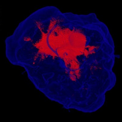
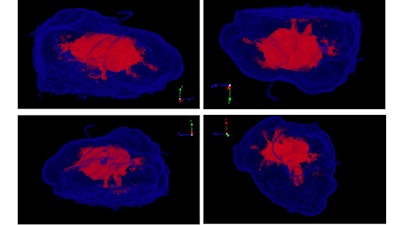 3D micro-CT visualization of breast tissue biopsy samples. Higher regions of x-ray absorption indicate cancer (red) and regions of lower x-ray absorption represent noncancerous tissue (translucent, blue). Image courtesy of James Michaelson, PhD.
After analyzing the 3D micro-CT data, the researchers found that physicians were able to detect cancerous tissue on the scans even when the cancerous tissue occupied only 0.45% of the surface area of the specimen. In comparison, the more conventional technique of examining pathology slides allowed clinicians to detect cancerous tissue only if it occupied at least 1.55% of the specimen surface area.
In all, 3D micro-CT detected 93% of the cancers deemed positive by pathologists, as well as 28 additional cases missed by the traditional method.
The 3D micro-CT technique has the potential to help physicians identify breast cancer margins not only more precisely than the histological method but also much more quickly, the authors found. The technique can help physicians identify the extent of cancer within minutes, compared with hours using the standard method, which could potentially enable surgeons to adjust and optimize their surgical approach while performing the operation.
"Today, one in three breast cancer patients goes home with cancer still in them," Michaelson said. "For most of these patients, this means returning for a second operation. For a small number of patients, this means local recurrence of the cancer left behind, resulting in about 2,000 deaths in the U.S. each year. 3D micro-CT has the potential to prevent most of this."

