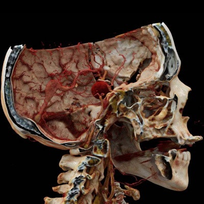
Cinematic rendering based on CT angiography (CTA) data can enhance the visualization of complex cerebrovascular anatomy and disease beyond the capabilities of traditional volume rendering, without any added costs, according to an article recently published online in the Journal of Neuroimaging.
Still relatively new to medicine, cinematic rendering continues to gain ground as a viable image-processing technique for enhancing the visualization of CT scans. Case studies have demonstrated its value in evaluating vascular anomalies and soft-tissue injuries and fractures, among other conditions.
The technique uses a global lighting model to produce photorealistic 3D image reconstructions with "greater nuance in surface texture and local shadowing, providing a richer sense of depth and spatial relationships," compared with existing reconstruction tools, noted Dr. Travis Caton and colleagues from Brigham and Women's Hospital (Journal of Neuroimaging, February 18, 2020). Caton is currently a diagnostic neuroradiology fellow at the University of California, San Francisco.
For the study, Caton led a team that performed cinematic rendering on the CT angiography data of patients with cerebrovascular diseases using volume rendering software (syngo.via, Siemens Healthineers).
After examining the cases under a variety of settings, the researchers identified several potential applications for cinematic rendering, which they divided into the following categories:
- Virtual anatomy lab: Cinematic rendering can serve as an adjunct educational tool to standard medical imaging or volume rendering in demonstrating complex cerebrovascular anatomic and pathologic relationships, the authors noted.
- Virtual physical exam: The high fidelity of cinematic rendering is capable of recreating the external appearance of soft tissues and skin with superior anatomic detail, compared with conventional imaging, they noted. This "lifelike" rendering of superficial anatomy, in conjunction with a physical exam, could enable clinicians to uncover clinical features of neurologic disease.
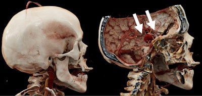
- Virtual craniotomy: Cinematic rendering images can also enhance preoperative planning for cerebrovascular procedures through a more heightened view of anatomy and pathology, compared with traditional volume rendering and CT angiography.
They used cinematic rendering to simulate cadaveric dissection at their institution's anatomy lab and found that the technique helped students, trainees, and subspecialty clinicians recognize certain aspects of cerebrovascular anatomy and pathology that are frequently missed on routine radiographic evaluation.
For example, clinicians were more easily able to identify thrombosis of the cortical veins and dural venous sinuses -- a clinically ambiguous disease often missed on routine imaging -- by examining cinematic rendering images, compared with conventional CTA scans.
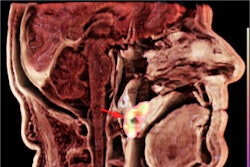
In one case, the researchers were able to use cinematic rendering to confirm the precise location of orbital varices not caught on the initial physical exam in a patient with intracerebral bleeding.
In effect, surgeons can virtually simulate cerebrovascular procedures at a workstation by repositioning and adjusting cinematic rendering images. Radiologists and neurologists may also use the technique to better understand the microanatomy of vascular malformations from the surgeon's perspective during preoperative planning.
 Cinematic rendering based on CT angiography scans of the head. "Virtual craniotomy" allows for removal of the calvarium and 3D visualization of the giant, surgically excluded middle cerebral artery aneurysm (white arrows). Image courtesy of Dr. Travis Caton.
Cinematic rendering "shows promise as a tool for clinical decision-making, teaching, and planning invasive procedures" and represents an emerging alternative to 3D printing, virtual reality, and augmented reality for personalized medicine, the authors wrote. The added advantage of cinematic rendering is that its use requires no additional devices or materials and is much less time-consuming than the other advanced visualization techniques.
"It is plausible that [cinematic rendering] will surpass or obviate the need for virtual and augmented reality systems and 3D printing for many current applications given these efficiencies," they wrote.


