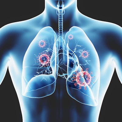
Artificial intelligence (AI) could enhance the role of chest imaging in the COVID-19 pandemic, moving it beyond disease evaluation to applications such as assessing risk, monitoring treatment, and discovering therapeutic targets, according to an editorial published May 6 in Radiology: Artificial Intelligence.
"AI's power to generate models from large volumes of information -- fusing molecular, clinical, epidemiological, and imaging data -- may accelerate solutions to detect, contain, and treat COVID-19," wrote a team led by Dr. Shinjini Kundu, PhD, of Johns Hopkins Hospital in Baltimore.
The standard test to determine infection with the SARS-CoV-2 virus is the reverse transcriptase polymerase chain reaction (RT-PCR) assay, but even this has been limited at times by a lack of tests and false negatives. Clinicians have used chest imaging, both x-ray and CT, as an additional tool to detect COVID-19 pneumonia, but both modalities have limitations, and professional groups such as the American College of Radiology (ACR) have warned specifically against using CT to diagnose the illness, recommending instead that it be used when "alternate diagnosis is suspected or when COVID-19 testing is unavailable or highly restricted," the authors wrote.
AI could help refine the use of chest imaging for COVID-19 applications, according to the group.
"Artificial intelligence (AI) could provide a stopgap while practicing radiologists understood and incorporated new findings into the diagnostic workflow," it wrote.
Kundu and colleagues described current research published on arXiv.org and medRxiv.org (neither of which perform peer review of submitted articles) that suggests AI's benefit when combined with chest imaging:
- An open-source, deep convolutional neural network called COVID-Net showed 83.5% accuracy in classifying normal, bacterial pneumonia, non-COVID viral pneumonia, and COVID-19 viral pneumonia on almost 6,000 posterior-anterior chest x-rays (Wang et al, arXiv, March 22).
- A 3D convolutional neural network showed 86.7% accuracy distinguishing COVID-19 pneumonia versus flu versus other disease on chest CT (Xu et al, arXiv, February 21). Another study using a system called DeepPneumonia found that the system could quickly find lesions and classify COVID-19 patients using chest CT data, with a reported area under the receiver operating curve of 0.99 (Song et al, medRxiv, February 25).
AI could expand chest imaging's role by helping clinicians predict prognosis of the disease, thus allowing for better care and treatment of COVID-19 patients. It could also help with the navigation of the "intricate link between lung pathology and immune inflammation in COVID-19 [as] a potential strategy to mitigate lung damage," according to the researchers.
"Coupled with a large and growing body of tools, AI has the potential to expand the role of chest imaging beyond diagnosis to facilitate risk stratification, treatment monitoring, and discovery of novel therapeutic targets in this global race to contain and treat COVID-19," they concluded.


