Measurements of muscle mass and density automatically extracted from chest CT exams using an artificial intelligence (AI)-based model can predict all-cause mortality over a six-year period in older men, according to research published online June 6 in the Journals of Gerontology.
A team of researchers led by Dr. Leon Lenchik of Wake Forest School of Medicine developed a three-stage automated approach for measuring baseline paraspinous skeletal muscle area (SMA) and skeletal muscle density (SMD) on chest CT images. Their goal was to find an alternative to dual-energy x-ray absorptiometry (DEXA) to derive measurements of muscle health as biomarkers for healthy aging.
After using the system to retrospectively calculate these metrics on patients from the U.S. National Lung Screening Trial (NLST), the authors found that higher levels of both muscle area and muscle density were associated with a statistically significant lower risk of all-cause mortality among older men. That wasn't the case, however, for older women.
Nonetheless, the study "confirms that CT-derived muscle metrics are useful prognostic biomarkers in older adults," the authors wrote.
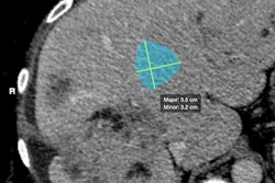
The researchers created a three-stage automated pipeline for skeletal muscle measurement using open-source machine-learning methods. After a random-forest classifier selects the appropriate chest CT series from the metadata on raw DICOM files, a convolutional neural network (CNN) then extracts the CT slices at the level of the T12 vertebra. In the third stage, another CNN segments the left paraspinous muscle from the single extracted CT image and then records the SMA and SMD, according to the researchers.
The entire pipeline was developed and tested using 2,084 CT exams from a subset of patients 70 years or older from the NLST. The researchers then applied their methods to the CT studies of 6,803 men and 4,558 women ages 60-69 from the NLST and used Cox proportional hazard models to assess the association between the muscle metrics and all-cause mortality.
They found that men with higher levels of muscle area and muscle density had a statistically significant lower risk of mortality as measured by hazard ratios. The differences were not statistically significant for women, however.
| Association between CT-derived muscle metrics and all-cause mortality in women and men ages 60-69 | ||
| Hazard ratio (HR) for all-cause mortality in women | Hazard ratio for all-cause mortality in men | |
| Skeletal muscle area (SMA) | 0.97 | 0.85* |
| Skeletal muscle density (SMD) | 0.98 | 0.91* |
Each standard deviation (3.5 cm²) increase in SMA in men was associated with a 17% decrease in mortality, while each standard deviation (6.99 Hounsfield units) increase in SMD was associated with an 11% decrease in mortality. The association of CT-derived muscle metrics with survival persisted after adjusting for age, race, smoking, cancer history, and other comorbidities, according to the researchers.
"Importantly, after adjusting for SMD (a measure of myosteatosis), SMA (a measure of muscle mass) remained predictive of mortality," the authors wrote. "Similarly, after adjusting for SMA, SMD remained predictive of survival. This indicates that muscle mass and myosteatosis contribute independently to survival in older adults."
The authors noted that this type of muscle screening could also be provided on chest CT scans for lung cancer screening.
"As the number of chest CTs performed in older adults increases, such evaluation of muscle metrics may help improve the cost-effectiveness of lung screening programs while at the same time providing valuable prognostic information about overall survival," the authors wrote.


