The less pulmonary consolidation seen on chest CT, the more likely COVID-19 patients will have had initial negative reverse transcription polymerase chain reaction (RT-PCR) test findings, according to a study published June 18 in the American Journal of Roentgenology.
The results confirm that chest CT plays a key role in thorough COVID-19 screening in patients who have negative RT-PCR tests but who still have symptoms of the disease, wrote a team led by Dr. Dandan Chen of Guangzhou First People's Hospital in Guangzhou, China. In fact, after the China National Health Commission released guidance in February on treatment of the novel coronavirus that described chest CT as a key tool for diagnosing COVID-19, use of the modality increased and more cases were identified.
"After this guidance was instituted, the number of new cases in Hubei ... increased ... which indicated that CT had a higher detection rate in patients with disease in the incubation period, especially for those with negative initial RT-PCR results," Chen and colleagues wrote. "This finding showed that CT is helpful for early diagnosis, timely isolation, and treatment of COVID-19 pneumonia."
Although RT-PCR is considered the gold standard method for diagnosing SARS-CoV-2 infection, it does have a vulnerability to false negatives, due to factors such as testing in early stages of COVID-19 pneumonia, insufficient cellular material gathered to test, varying test methods, and operator skill. Chen and colleagues sought to explore whether CT could clarify COVID-19 diagnoses in patients with negative RT-PCR results but disease symptoms.
Their study included 21 patients with (eventually) confirmed COVID-19 treated at five hospitals in Guangzhou between January 19 and February 20. Each patient underwent chest CT and RT-PCR testing. The researchers divided the patients into two groups: one group of seven with initial negative RT-PCR results (found to have positive results after the test was repeated two days later), and another group of 14 patients with initial positive RT-PCR test results.
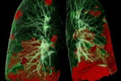
Across the patient cohort, the group noted the following findings on chest CT:
- 67% of COVID-19 lesions were found in multiple lobes.
- 72% of COVID-19 lesions were found in both lungs.
- 95% of CT COVID-19 findings showed ground-glass opacities, while 72% showed consolidation.
- 33% of patients had other lesions around the bronchovascular bundle.
- 57% of patients had air bronchogram and 67% had vascular enlargement.
- 62% had interlobular septal thickening and 19% had pleural effusions.
However, when Chen's team compared the patients who had initial negative results with those who had initial positive RT-PCR results, the researchers found that chest CT scans in the negative results group were less likely to show pulmonary consolidation (three patients out of seven, compared with 12 patients out of 14; p < 0.05).
Although CT shouldn't be considered the be-all and end-all for diagnosing COVID-19, it is an important tool, according to the investigators.
"When patients with suspected COVID-19 pneumonia who have an epidemiologic history and typical CT features have negative initial RT-PCR results, repeated RT-PCR tests and patient isolation should be considered," they concluded.


