CT has a key role to play in the diagnosis of COVID-19 pneumonia, according to a July 16 session at the virtual 2021 Society of Cardiovascular Computed Tomography (SCCT) meeting.
Presenters at a session titled "Cardiovascular imaging during the COVID-19 pandemic" described how CT can not only help stratify patients' complication risk but also improve their care when, say, incidental COVID-19 lung findings are identified on cardiac CT.
Dr. Diana Litmanovich of Harvard University in Boston kicked off the discussion by outlining the advantages of cardiac CT and coronary CT angiography (CCTA) -- which have proven to be effective tools for classifying patients' risk for complications from COVID-19.
"[For example,] coronary CTA might play an important role in diagnosis, triaging, and risk stratification [for myocarditis] in acute cases," she said.
Litmanovich cited guidance from a May 2020 statement published in the International Journal of Cardiovascular Imaging on ways to reduce scarcity of healthcare resources during the COVID-19. She noted that CT offers an alternative to more invasive tests, such as coronary angiography for further workup of elevated troponin, diagnostic of uncertain non-ST segment elevation myocardial infarction (NSTEMI), acute chest pain, transesophageal echocardiogram for transcatheter aortic valve replacement or surgical aortic valve replacement planning.
"CCT can be used to minimize risk [from other more invasive imaging modalities], reduce resource utilization, and maximize clinical benefit," she said. "And it has the added advantage of evaluating lung parenchyma, pulmonary embolism, and right ventricular failure."
Later in the session, Dr. J. Jeffrey Carr of Vanderbilt University in Nashville, TN, offered a primer of how incidental COVID-19 findings may appear on cardiac CT and coronary artery calcium (CAC) scoring exams.
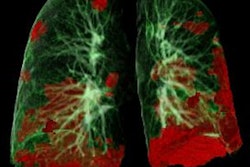
"The most commonly reported CT findings in COVID-19 patients are typical of an organizing pneumonia pattern of lung injury," he said. "[In fact], early COVID-19 respiratory disease is better understood primarily as SARS-CoV-2-induced secondary organizing pneumonia."
COVID-19 pneumonia will make itself known on cardiac CT, and identifying it improves patient care, Carr said. Signs include typical round peripheral, crazy-paving pattern, and ground-glass opacities; pulmonary emboli and "other thrombotic complications" are common as well, he concluded.


