An artificial intelligence (AI) algorithm can highlight incidental cases of pulmonary embolism (PE) on contrast-enhanced chest CT exams that were performed for reasons other than to detect PE, according to a presentation at the recent online annual conference of the Society for Imaging Informatics in Medicine (SIIM).
A team of researchers from Yale University retrospectively evaluated a machine-learning algorithm on chest CT exams performed at their institution over a two-month period. They found the model to be extremely sensitive and specific.
"This algorithm could help the radiologist detect incidental PEs, it could expedite reporting of cases with incidental PEs, and can help identify patients that may need immediate medical attention," said first author and presenter Dr. Anna Bader.
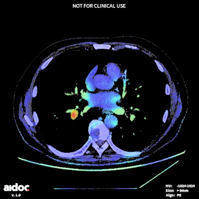
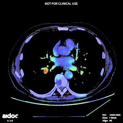 Color map of CT image flagged by AI software as a positive study for PE. The 59-year-old man, who was undergoing pancreatic cancer restaging, was found to have a segmental PE. Image courtesy of Dr. Anna Bader.
To assess its performance, the researchers retrospectively applied Aidoc's PE model to 2,632 consecutive contrast-enhanced chest CT studies from January 1, 2018, to February 28, 2018. CT pulmonary angiograms were not included.
After natural language processing (NLP) was utilized to classify the radiologists' reports, two board-certified thoracic radiologists reviewed all cases that had discrepant results between the algorithm and the radiology reports. In cases where the two radiologists differed, a third thoracic radiologist served as the tiebreaker.
The algorithm yielded extremely high accuracy and performance for the 34 PE cases (1.3% of chest CT exams) that were found in the study, Bader said.
| Performance of machine-learning algorithm for incidental detection of PE | |
| Accuracy | 99.5% |
| Sensitivity | 94.1% |
| Specificity | 99.6% |
| Positive predictive value | 74.4% |
| Negative predictive value | 99.9% |
The researchers found 28 discrepant results between the original radiologist report and AI algorithm. Of these, 25 were positive on AI but negative on the report. There were three cases that were negative on AI and positive on the report.
Upon further review by the thoracic radiologists, 14 of the 25 cases that were positive on AI and negative on the report were deemed to be true-positive cases. These included six segmental, six subsegmental, and two lobar PEs. None of these patients demonstrated right heart strain on CT, she said.
Of the three cases that were negative on AI but positive on the report, two were positive on expert review. One was segmental and one was subsegmental, according to Bader. Again, none of these patients had right heart strain.
"This machine-learning algorithm is extremely sensitive and extremely specific," she said. "The very, very high negative predictive value makes this a great screening method."
The algorithm is already live at their institution, she said.
"Future work with his algorithm will help to determine whether there's an improved detection rate when the radiologist uses it in the course of their workflow," she said. "It will [also] be useful to assess if there's a decrease in turnaround time for positive cases that are flagged by the algorithm. Finally, and possibly most importantly, would be to determine whether there's an outcome benefit for patients in the detection of these mostly small pulmonary emboli."


