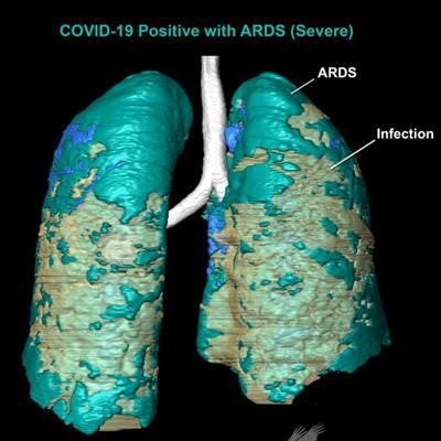
Can advanced visualization techniques used to research the lungs of reptiles and birds also help diagnose COVID-19 in humans? Applying methods from evolutionary anatomy research, a Louisiana State University (LSU) team has created 3D digitally segmented models that demonstrate the extent and distribution of the disease.
Not only that, but these models could be 3D-printed and used to communicate the impact of the disease to members of the public, according to co-authors Emma Schachner, PhD, an evolutionary anatomist, and Dr. Bradley Spieler, a radiologist at LSU Health Sciences Center New Orleans.
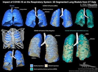
In an article published online August 18 in BMJ Case Reports, the researchers shared 3D digital models that were created from lung CT exams of three patients who had been hospitalized with worsening symptoms associated with SARS-CoV-2.
"Three-dimensional segmented digital models provide a dramatically clearer method for visually evaluating the impact of COVID-19 on the lungs than straight radiographs, CT data, or reverse transcription polymerase chain reaction alone," the authors wrote. "These printable digital models are additionally very powerful for communicating the impact of COVID-19 on the respiratory system to the general public."
3D surface models were segmented manually from contrast-enhanced chest CT exams using the Avizo 7.1 scientific visualization software (Thermo Fisher Scientific) and methods utilized before by the institution for evolutionary anatomy research. Thin-section (1 mm) CT datasets were used to produce the models.
"Previously published 3D models of lungs with COVID-19 have been created using automated volume rendering techniques," Schachner said in a statement from LSU. "Our method is more challenging and time consuming, but results in a highly accurate and detailed anatomical model where the layers can be pulled apart, volumes quantified, and it can be 3D printed."
 3D-segmented models of lung CT data show the distribution of COVID-19-related infection in the respiratory system. Image courtesy of LSU Health New Orleans.
Two of the three patients had positive reverse transcription polymerase chain reaction (RT-PCR) results for SARS-CoV-2, but the third case was negative. This patient was considered to be a false-negative result, however, due to compelling clinical and imaging features indicative of COVID-19, according to the researchers.
Given the diagnostic challenges related to false-negative RT-PCR results, CT can be helpful in establishing a COVID-19 diagnosis, Schachner and Spieler said.
"Importantly, these CT features can range in morphology and appear to correlate temporally with disease progression," they wrote. "This allows for 3D segmentation of the data in which lung tissue can be volumetrically quantified, or airflow patterns could be modelled."
What's more, the holistic view provide by these 3D models makes it easier for a wider audience to appreciate the extent of pulmonary disease, according to the researchers.
"Unlike simple volume rendered images, these models can be 3D printed, and thus have a much broader functional application that allows for the collaboration between basic and clinical scientists, which is particularly important given the critical nature of COVID-19," they wrote.
Schachner and Spieler said they are now segmenting additional models for a larger follow-up project.


